Control Device Thermal Modeling in MRI Receive Coil Q-Spoiling Circuits
Robert H. Caverly, Savannah Benbrook
Read Description
These semiconductor devices can see the full RF power during operation, and will exhibit significant heating, potentially leading to device failure. Even if the device does not reach a destructive heating level, the resistance of the device can increase and effectively degrade the level of blocking in the RF coil. A useful tool for MRI circuit designers would be a thermal model that predicts this change in resistance, and hence degradation in MRI coil blocking, as device temperature rises. This paper presents a method for modeling thermal effects in MRI Q-spoiling control devices. A PIN diode resistance-temperature model is reviewed, and two approaches for field effect transistors that model the temperature rise and subsequent device resistance variation with temperature using SPICE-compatible equivalent circuit elements are covered. Experimental results are presented that validate the form of the models and show that increased device resistance due to temperature rise will degrade MRI blocking by several dB. The ability to model the control device’s thermal characteristics will lead to improved understanding of circuit operation.
×
Take Out Message
Take-Home Messages
- The level of MRI coil detuning and blocking is strongly dependent on the resistance of the control device which can change due to device heating
- More robust circuitry and understanding of both the electrical and thermal operation of the devices (and their interplay) will lead to more reliable MR instrumentation.
- The targeted medical application is MRI receive coil detuning/blocking circuits to protect the patient and the sensitive electronics in the presence of the high power slice-select pulse.
- The ability to model the control device’s thermal characteristics will lead to improved understanding of circuit operation, aid in the selection of control device for the specific MR application, and will lead to more reliable MRI instrumentation.
Image Registration for Microwave Tomography of the Breast Using Priors from Non-Simultaneous Previous Magnetic Resonance Imaging
Gregory Boverman, Cynthia Davis, Shireen Geimer
Read Description
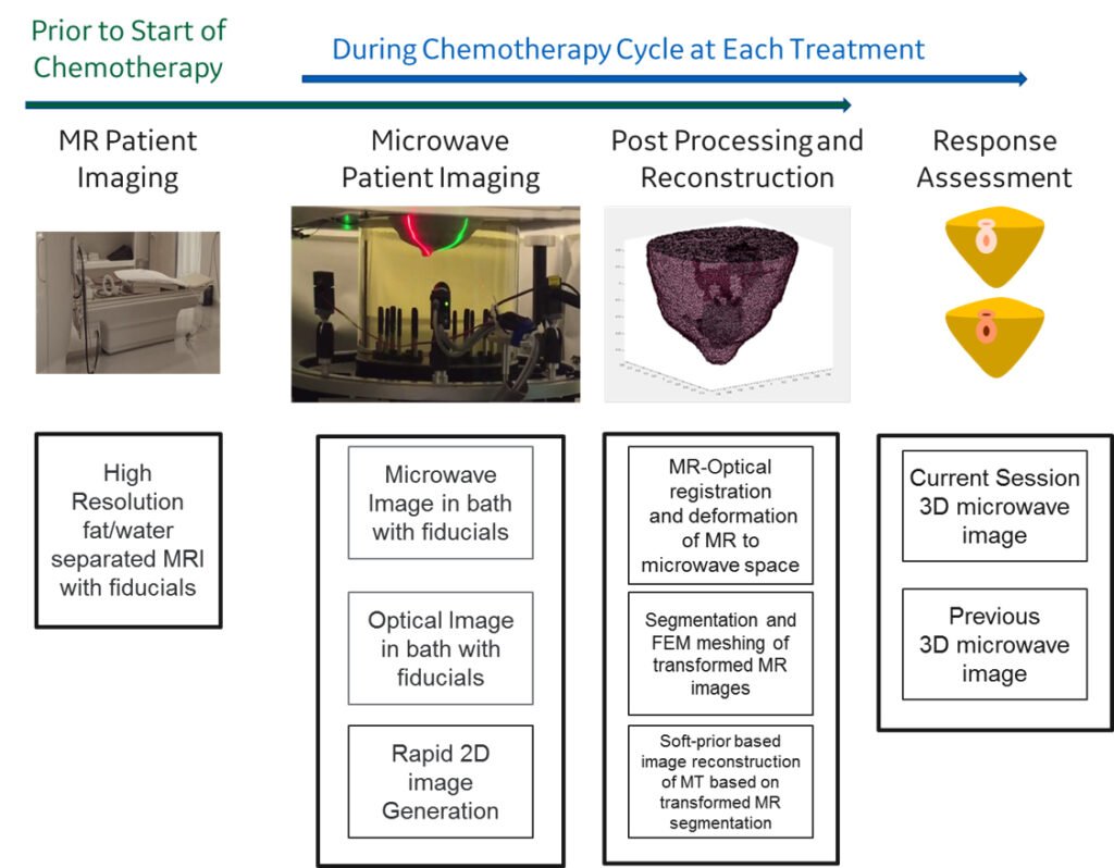
For the neoadjuvant therapy application, it would be desirable to achieve the most accurate possible characterization of the tissue properties. One method to achieve increased resolution and specificity in microwave imaging reconstruction is the use of a soft prior regularization. The objective of this study is to develop a method to use magnetic resonance (MR) images, taken in a different imaging configuration, as this soft prior. To enable the use of the MR images as a soft prior, it is necessary to register the MR images to the microwave imaging space. Registration fiducials were placed around the breast that are visible in both the MRI and with an optical scanner integrated into the microwave system. Utilizing these common registration locations, numerical algorithms have been developed to warp the original breast MR images into a geometry closely resembling that in which the breast is pendant in the microwave system.
×
Take Out Message
Take-Home Messages
- To achieve increased resolution and specificity in microwave imaging reconstruction a soft prior regularization can be used from pre-microwave imaging MRI performed as part of standard clinical care for breast cancer.
- The targeted application is breast cancer imaging for neoadjuvant chemotherapy monitoring.
- Utilizing a limited set of skin fiducial markers numerical algorithms have been developed to warp the original breast MR images into a geometry closely resembling that for when the breast is pendant in the microwave system.
- Leveraging magnetic resonance images acquired prior to neoadjuvant chemotherapy a soft prior regularization of the microwave imaging can be performed to increase resolution and specificity thus potentially improving the ability of the oncologist to predict chemotherapy effectiveness.
A First Evaluation of the Realistic Supelec-Breast Phantom
Tomas Rydholm, Andreas Fhager, Mikael Persson
Read Description
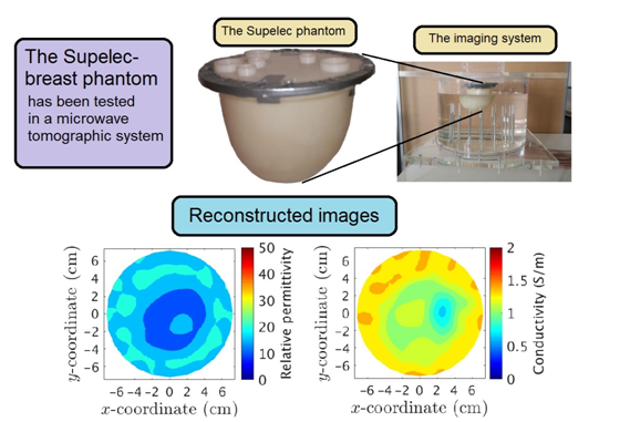
An iterative algorithm is then used to solve the inverse problem and reconstruct a 2-D plane transecting the phantom. The reconstructed images are compared to the ones recovered from a cylindrical phantom of equivalent phantom media. The results show that both phantoms are possible to reconstruct, although the interior of the Supelec phantom is more challenging.
×
Take Out Message
Take-Home Messages
- We have used our microwave-imaging system in order to successfully reconstruct the Supelec-breast phantom and evaluate its usability.
- Our system is capable of recover images of the studied phantoms, although the interior of the Supelec phantom is challenging due to the high plastic content.
- The studied phantom was developed to be a tool within the microwave-imaging community.
- These are the first published reconstructed images of the Supelec phantom and shows that the phantom is a useful tool when developing a microwave-imaging system.
- It is also shown that our system is capable of reconstructing an image of the phantom without utilizing anya prioriinformation of the phantom, which is unique in the microwave-imaging community.
A Novel Approach for Determining the Electromagnetic Properties of a Colloidal Fluid with Magnetic Nanoparticles for Hyperthermia Applications
Danilo Brizi, Nunzia Fontana, Giulio Giovannetti, Alessandra Flori, Luca Menichetti,Saer Doumett, Giovanni Baldi, Agostino Monorchio,
Read Description
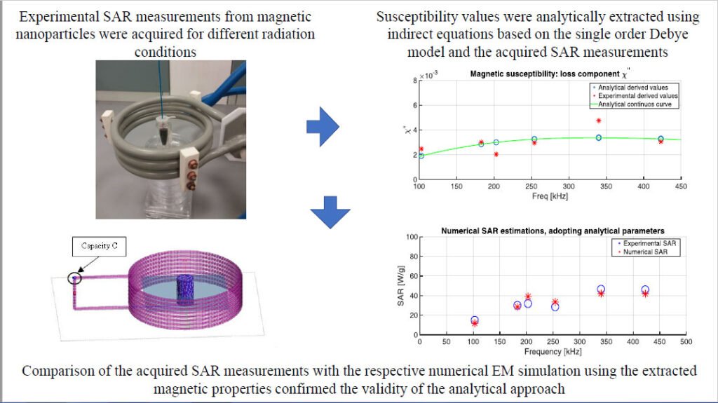
In particular, it makes use of indirect equations based on the single order Debye model combined with a punctual set of in vitro SAR measurements. The procedure has a general validity and it can be easily applied in the up-growing field of magnetic hyperthermia studies.
×
Take Out Message
Take-Home Messages
- A novel approach able to deeply optimize clinical treatments of magnetic hyperthermia with nanoparticles.
- Innovative determination of the complex magnetic permeability of magnetic colloidal fluid by simply using invitro SAR measurements.
- A rapid, alternative and broadband approach for determining the electromagnetic properties of magneticnanoparticles.
- Possibility to test the efficiency of nanoparticles in a tissue-like environment: optimized and realistictreatment planning for magnetic hyperthermia.
Dual-Band Skin-Adhesive Repeater Antenna for Continuous Body Signals Monitoring
Joao Felicio, Carlos A. Fernandes, Jorge R. Costa
Read Description
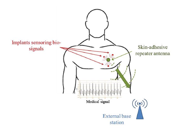
The proposed adhesive repeater is low profile, and it is designed for dual-band operation at the ISM band (2.4 GHz) and in the ultrawideband (UWB) spectrum (4-10.6 GHz) using a single excitation port. The ISM band is used for in-body communication with the implants and the UWB band for off-body burst communication with the BS. The antenna consists of three layers that grant it compactness and performance robustness. We assess the in-body link between the repeater and a custom-designed miniaturized implantable probe antenna. The study is performed in the frequency-domain with results showing adequate input impedance matching (s11 ≤ -10 dB) and robustness to different body parts. We extend the analysis to time-domain by transmitting a synthetized medical signal between the two antennas. In addition, we evaluate the feasibility of an off-body link for burst communication. Again, the results indicate very good performance by the repeater antenna both in terms of impedance matching (s11 ≤ -10 dB) and preservation of time-signals (fidelity ≥ 75%), which is relevant in impulse radio systems. To the authors’ best knowledge, this is the first time this kind of dual-band adhesive antenna is being proposed for BANs, complete with performance tests using frequency- and time-domain figures-of-merit.
×
Take Out Message
Take-Home Messages
- We assess the viability of having a dual-band skin-attached repeater antenna for in- and off-body communication for the transmission of medical data in the context of Body Area Networks. It is the first time a single-port antenna with both ISM (2.4 GHz) and UWB (4-10.6 GHz) operation bands is proposed for BAN applications.
- The study demonstrates the implementation of a skin-adhesive antenna and the feasibility of both in- and off-body communication links with a passive implant and an external base station, respectively.
- The proposed repeater antenna can be used in continuous monitoring applications, in order to detect health conditions in advance.
- We study for the first time the possibility of having a skin-attached repeater antenna with capability of establishing an in-body communication link with multiple implants and further relay the collected information through a UWB off-body link, thus increasing the off-body communication range and also diminishing the transmission time.
- The present paper includes the complete study of the in- and off-body links not only in the time-domain, in which we transmit a QPSK-modulated ECG signal, but also in the frequency-domain, where we assess the power transfer and link budget.
Channel Characteristics and Wireless Telemetry Performance of Transplanted Organ Monitoring System Using Ultra-wideband Communication
Pongphan Leelatien, Koichi Ito, Kazuyuki Saito, Manmohan Sharma, Akram Alomainy
Read Description
Ultra-wideband technology is well suited for wireless implanted applications since it allows physical size reduction as well as increased longevity of the implanted devices. However, because of the high attenuation of UWB signals inside the human body, it is necessary to examine their propagation characteristics and demonstrate the feasibility of such application. In this paper, an investigative study involving the liver implanted wireless telemetry link using UWB technology is presented. Measurements using multilayer phantoms and simulations using a human digital model have been conducted within the frequency band of 4.5-6.5 GHz. Multilayer phantom measurements have demonstrated the attenuations ranging between -50 dB and -100 dB over the considered frequency band. A path loss model at the liver area has been obtained from the simulations. Also, the numerical results have shown that the attenuation variation due to respiration-induced organ movements was within 30 dB range with respect to the largest organ movement distance of 40 mm which emphasized the influence of organ movements on the in-body attenuations. Our preliminary link budget evaluation of liver-skin surface communication link including the effect of shadowing and organ movements estimated that it is possible to achieve a high data rate of 10 Mbps with a bit error rate of 10−3 at a distance of about 40 mm.
×
Take Out Message
Take-Home Messages
- This application utilizes wireless communication to transmit biological data from the transplanted liver to provide constant and real-time monitoring after transplantation.
- This work shows that it is feasible to establish wireless communication from the liver area to the body surface and thus to implement such intended applications in the described scenario using ultra-wideband (UWB) technology.
- The potential medical applications include wireless monitoring and diagnostics of internal organs of the human body as well as drug delivery at specific locations.
- This work offers, for the first time, the fundamental understanding of UWB channel characteristics at the liver location considering respiration-induced organ movements.
- With the potential utilization of ultra-wideband technology for liver implanted wireless communications, this would enable other future innovative healthcare applications.