Real-time Microwave Imaging of a Compressed Breast Phantom with Planar Scanning
Daniel Tajik, Farzad Foroutan, Denys S. Shumakov, Aaron D. Pitcher, Natalia K. Nikolova
Read Description
Despite the fact that these algorithms are based on a linear forward model of scattering, they have been capable of providing quantitative estimates of the tissue permittivity due to the experimentally derived kernel of the scattering integral. Here, we demonstrate similar performance with a thicker (about 5 cm) compressed-breast phantom. This thickness is greater or comparable to the median thickness employed in mammography, depending on the view (craniocaudal or mediolateral oblique). The two methods are described in a common mathematical framework for the first time. The importance of the system calibration and the choice of a host medium are discussed through experiments. A new method for focusing onto suspect regions is demonstrated. The limitations of real-time imaging are highlighted along with an outlook to improving the image resolution and suppressing artifacts without sacrificing the reconstruction speed.
×
Take Out Message
Take-Home Messages
Quantification of the Sensing Radius of a Coaxial Probe for Accurate Interpretation of Heterogeneous Tissue Dielectric Data
Alessandra La Gioia, Saqib Salahuddin, Martin O’Halloran, Emily Porter
Read Description
Generally, uncertainties are higher in the dielectric measurement of heterogeneous tissues, due to the fact that there is no standard procedure for acquiring and interpreting the dielectric data of heterogeneous tissues. Uncertainties related to tissue heterogeneity can be minimised by estimating the probe sensing volume, defined by the sensing depth and radius, and characterising the tissue distribution within that volume. While several studies have investigated the sensing depth, this work focuses on examining the sensing radius. Both dielectric measurements and numerical simulations with heterogeneous porcine tissues in the microwave range of 0.5-20 GHz have been conducted to quantify the sensing radius and the dielectric contribution of each tissue within the sensing volume. Experiments demonstrate that the sensing radius, which depends on the individual dielectric properties of the constituent tissue types, can be smaller than the probe radius. This work further quantitatively demonstrates that the dielectric contribution of a particular tissue depends on both its location within the sensing volume and its dielectric properties. This study provides fundamental knowledge for accurately interpreting dielectric data of heterogeneous tissues, with the aim of supporting medical device development.
×
Take Out Message
Take-Home Messages
- This work analyses the sensing radius of a coaxial probe for accurate dielectric characterisation of heterogeneous tissues.
- The probe sensing radius can be smaller than the probe radius and depends on the histology of the tissue sample.
- Accurate knowledge of the sensing radius has the potential for improving the design of novel microwave imaging devices and hyperthermia systems.
- This work demonstrates that a lack of knowledge of the probe sensing radius leads to errors in the interpretation of dielectric data acquired from heterogeneous tissues, and thereby to inaccurate medical device design.
- Despite the assumption made in previous dielectric studies, this work shows that the dielectric contribution of a particular tissue depends on both its location within the sensing volume and its dielectric properties.
Magnetic Transceiver Beamforming for A 2 × 2 Magnetic Resonance Charging System
Xiaoqing Liu, Bingqing Mei, Xiaodong Wang, Zhigang Wen
Read Description
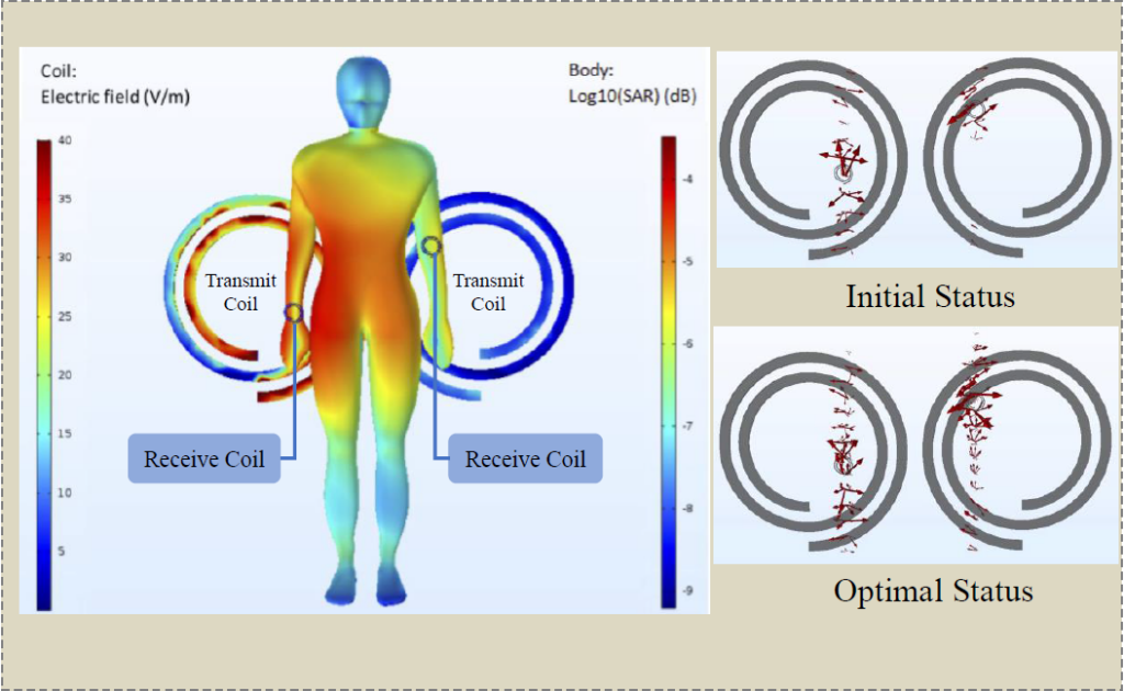
The formulated problem is nonconvex and we propose an efficient iterative solution by solving a series of geometric programs that approximate the original problem. Both numerical results and simulation results in COMSOL model are provided to demonstrate the effectiveness of the proposed biomedical wearable system and optimization algorithm.
×
Take Out Message
Take-Home Messages
- This paper proposes a 2×2 magnetic resonance charging system considering both magnetic transmit beamforming and receive beamforming for charging wearable medical devices.
- Both numerical results and simulation results in COMSOL model are provided to demonstrate the effectiveness of the proposed system and optimization algorithm.
- The target medical applications are wearable medical devices using for Alzheimer’s disease, ergonomics, rehabilitation and neurology which are inconvenient or sometimes infeasible to replace or change batteries for the elderly or patients.
- Such a magnetic resonance charging system provides a promising way to make wearable medical devices permanently unplugged.
Transmission Line Model Of An Implemented Insulated Cable For Magnetic Resonance Imaging Radiofrequency Hazard Evaluation
Alexia Missoffe, Julie Kabil, IADI, Pierre-Andre Vuissoz, Jacques Felblinger
Read Description
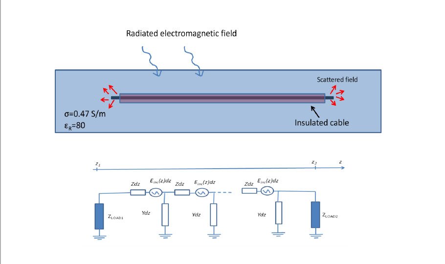
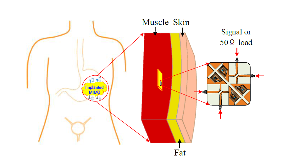
The equivalence between the models is shown through a finite difference model and a new analytical formula for the transfer function as a function of transmission line parameters. First, the possibility of modeling an insulated cable with a transmission line model was analyzed through full-wave numerical simulations. The assumption of a transmission line model underlying the transfer function model was shown to be right for a simple cable embedded in tissue imitating gel and the transmission line parameters extracted from this analysis were consistent with analytical formulas derived from the laws of physics. The transmission line model predictions were first compared to experimental and simulated data of the cable transfer function and then, to experimental and simulated data of the resonant behavior as a function of length of cables with different termination conditions. The measured and simulated transfer functions fit perfectly a transmission line model with an analytical expression of the propagating constant. The transmission line model extracted from the transfer function allows to predict the resonant behavior of two cables with different termination conditions
×
Take Out Message
Take-Home Messages
- An insulated cable implanted in a lossy material and submitted to a radiated radiofrequency field can be modeled by a modified transmission line with a distributed excitation along the line.
- This model gives a better physical insight into the problem of the compatibility of implanted cables in Magnetic Resonance Imaging (MRI) such as pacemaker, defibrillator or neurostimulator leads.
- This work shows the equivalence between the transfer function model usually used to model the interaction of an implanted lead with the radiofrequency field of MRI and the modified transmission line model.
- An idea that can be derived from this model is to reduce the heating at the electrode of an implanted lead by creating a big reflection coefficient before the end of the lead using a multi-section approach
Magnetoresistive Biosensors for On-Chip Detection and Localisation of Paramagnetic Particles
Zhaochen Yin, Edoardo Bonizzoni, Hadi Heidari
Read Description
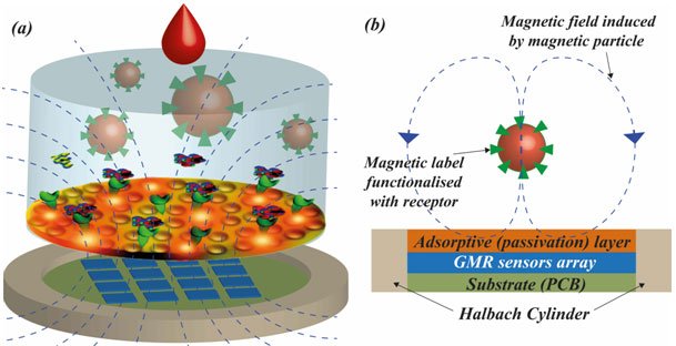
A new calibration and localisation algorithm has been coded and implemented. In order to generate the required homogenous magnetic field, a custom 3D printed Hallbach cylinder has been simulated and characterised. The system includes sensory and electronic boards to collect the data and to transfer them to a computing server. The experimental results are displayed in a visual interface. Ferrofluid is used to model and simulate the magnetic field change of the cell. This paper demonstrates a 4×4 sensors array and provides a step towards the miniaturised on-chip magnetoresistive based cell detection and localisation for portable diagnostics applications.
×
Take Out Message
Take-Home Messages
- This paper presents the design and the implementation of an on-chip magnetoresistive sensors array for cell detection and localisation. Giant magnetoresistance (GMR) sensors have been used due to their high sensitivity and resolution.
- A novel calibration and localisation algorithm has been coded and implemented. In order to generate the required homogenous magnetic field, a custom 3D printed Hallbach cylinder has been simulated and characterised.
- Sensory chips could detect an average magnetic sensitivity of 2 V/T at room temperature.
- The ferrofluid and a customised 3D printed Halbach cylinder were employed to simulate the magnetic field change in the cell.
- The implemented algorithm helps in achieving high sensitivity and positioning speed, also thanks to an accurate calibration of the GMR sensors.
Characterisation of the Dielectric Properties of the Bladder over the Microwave Range
Emily Porter, Saqib Salahuddin, Alessandra La Gioia, Adnan Elahi, Atif Shahzad, Arun Kumar, David Kilroy, M. O’Halloran,
Read Description
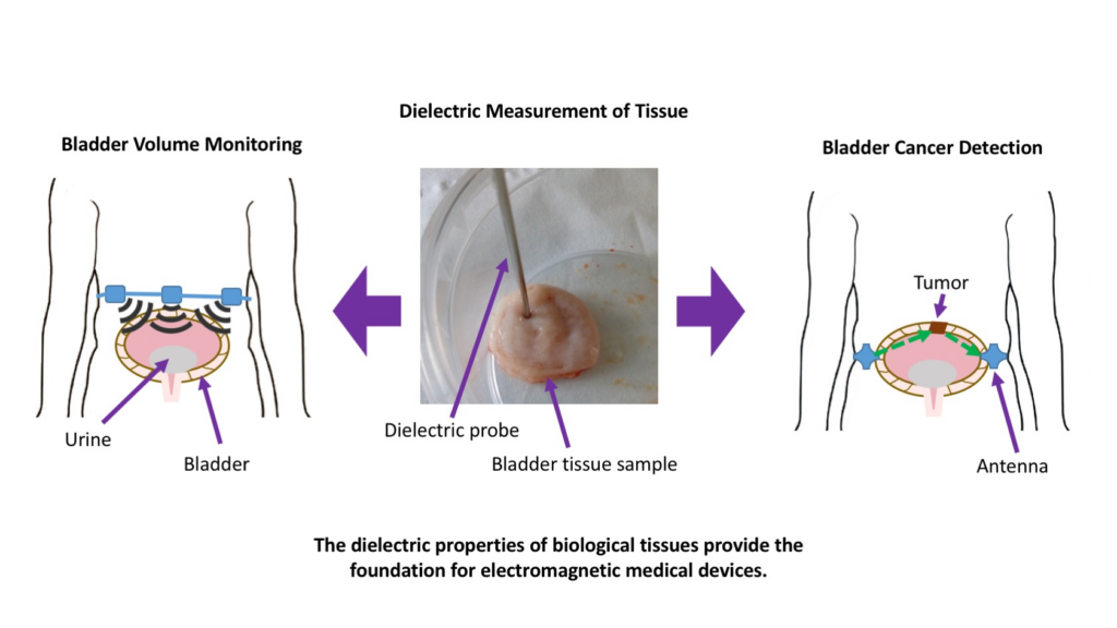
A number of such technologies have been proposed to target the diagnosis or treatment of bladder conditions, including urinary incontinence and bladder cancer. However, available dielectric data for the bladder is extremely limited in scope and has not been updated in line with modern knowledge, including best practices in measurement procedures to reduce the impact of confounders, along with improved reporting of experimental metadata. For these reasons, in this work we present a study of dielectric measurements on the bladder of freshly excised bovine and porcine tissues over the microwave frequency range. We examine the properties of the inside and outside of the bladder wall, and carefully control and record confounders during the measurements. The obtained data is analysed in terms of confounders (temperature, time from excision, inter-species differences), and compared to data from the literature. The resulting dataset, composed of 52 measurements from 10 animals, provides a thorough and quantitative understanding of the dielectric properties of the bladder. The results of this study will benefit the development of related electromagnetic medical technologies, and thereby positively impact on patient care over the long-term.
×
Take Out Message
Take-Home Messages
- Accurate knowledge of the dielectric properties of tissues is the basis for electromagnetic (EM) medical device design, development, and optimisation, and therefore, lack of knowledge on the dielectric properties of the bladder is a stumbling block for the application of bladder-related technologies, including EM therapeutic and diagnostic tools.
- This study provides a critical, anatomy-informed dielectric study of the properties of the bladder.
- Through a large number of tissue measurements, this study examines for the first time the impact of the dielectric properties of the bladder in light of common measurement confounders (time from excision, temperature), and uniquely examines the properties of both the inside and the outside of the bladder wall.
- This study provides reliable dielectric data of the bladder over the microwave frequency range, and fitted dielectric models, that can be used to support the design of bladder-related medical technologies.
- This study has been performed in line with modern best practices standards for the reporting of experimental metadata along with experimental data, and the data and metadata will be made openly available online.