Power Reduced Monolithic Wireless Sensor
Read Description
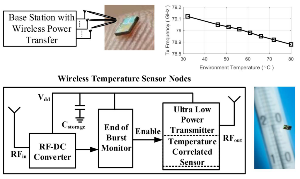
Combining highly-integrated ultra-low-power millimeter wave (mm-wave) sensing, wireless power transfer (WPT) and on-chip antenna is a path towards battery-less, fully monolithically integrated, millimeter-sized sensor nodes with only a few milligram of weight. In this work, a passive fully integrated monolithic wireless sensor for mm-wave sensing is presented in a 65 nm CMOS technology, including an on-chip wireless power receiver, on-chip antenna, and a low power transmitter.
Near Field Wireless Power Transfer to Stent-Based Biomedical Implants
Read Description
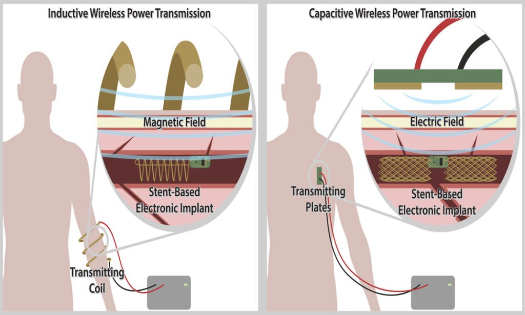
Moreover, stent-based devices are being recognized as a minimally invasive alternative to traditional surgery. Hence, the idea of using the body of the stent as the power receiving element is becoming increasingly attractive. The objective of this work is to analyze two near field wireless power transfer methods to stent-based devices, viz., inductive and capacitive coupling. The methods used are lumped element modelling, ac circuit theory, finite-element analysis and experiments to validate the model with excised bovine muscle tissue. Capacitive coupling is proposed as an alternate method due to the transmitter design that can be worn anywhere on the body. It achieves power transfer efficiencies of 2.6% and 1% when placed at depths in muscle tissue of 15 mm and 30 mm respectively. Safety requirements are also met. The capacitive link can accept an input power of 53 mW before exceeding the safe specific absorption rate limit of 1.6 W/kg averaged over 1 g of tissue.
A Fully Integrated Low-Power 30 GHz Complex Dielectric Sensor in a 0.25-μm BiCMOS Technology
Read Description
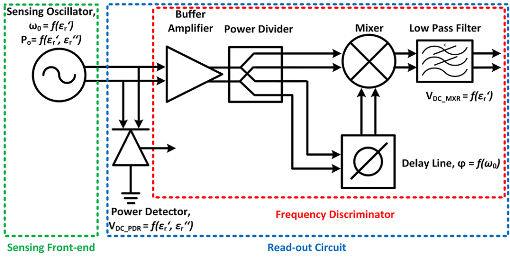
The MUT is exposed on the resonator component in a sensing oscillator and the oscillator results in permittivity and conductivity dependent change in the output frequency and output power, respectively. The frequency information is translated into DC voltage using a frequency discriminator and the output power is detected using a power detector. The sensor has been calibrated using iso-propanol, ethanediol and acetone solutions. Methanol-ethanol mixture solutions, in steps of 25% of concentration change, have been used to demonstrate the functionality of the sensor. The selectivity is showed using methanol-ethanol mixtures with concentration differences of 5% around a mixture ratio of 50:50. The chip is 2.3 sq. mm in size and consumes 60 mW power. The sensor measures complex permittivity within 3.7% accuracy for the real part and 4.3% for the imaginary part. As a compact and low-power solution the sensor is a potential candidate for minimal invasive investigations of chemicals and bio-materials at mm-wave frequencies.
Solutions to improve the outcomes of thermal treatments in oncology: multi-point temperature monitoring
Read Description
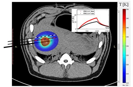
liver, lung, and prostate). In this context, the knowledge of temperature during treatment may be useful to better control the amount of damaged tissue and to subsequently improve clinical outcomes. The objective of this work is to assess the feasibility of two multi-point probes for temperature monitoring during laser ablation. The probes consist of a needle made up of a carbon fiber tube. Each probe embeds an array of 7 fiber Bragg grating sensors. Experiments performed in in vivo animal models (pig livers) show that the probe can reach deep-seated organs and offer the possibility to monitor tissue temperature in seven different positions. This information may be crucial to guide clinicians in the optimization of treatment settings and to improve the accuracy of theoretical models which will pilot future studies to design new heating devices and to develop patient-specific treatments
Real-time Microwave Imaging of a Compressed Breast Phantom with Planar Scanning
Read Description
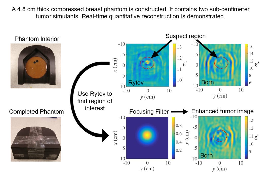
Despite the fact that these algorithms are based on a linear forward model of scattering, they have been capable of providing quantitative estimates of the tissue permittivity due to the experimentally derived kernel of the scattering integral. Here, we demonstrate similar performance with a thicker (about 5 cm) compressed-breast phantom. This thickness is greater or comparable to the median thickness employed in mammography, depending on the view (craniocaudal or mediolateral oblique). The two methods are described in a common mathematical framework for the first time. The importance of the system calibration and the choice of a host medium are discussed through experiments. A new method for focusing onto suspect regions is demonstrated. The limitations of real-time imaging are highlighted along with an outlook to improving the image resolution and suppressing artifacts without sacrificing the reconstruction speed.
Quantification of the Sensing Radius of a Coaxial Probe for Accurate Interpretation of Heterogeneous Tissue Dielectric Data
Read Description
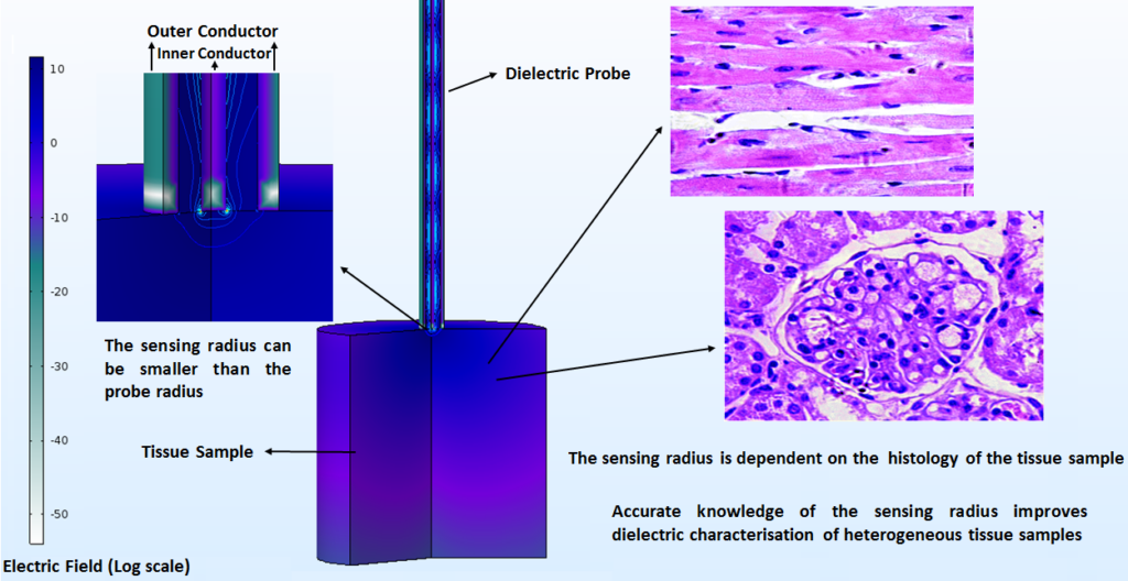
Generally, uncertainties are higher in the dielectric measurement of heterogeneous tissues, due to the fact that there is no standard procedure for acquiring and interpreting the dielectric data of heterogeneous tissues. Uncertainties related to tissue heterogeneity can be minimised by estimating the probe sensing volume, defined by the sensing depth and radius, and characterising the tissue distribution within that volume. While several studies have investigated the sensing depth, this work focuses on examining the sensing radius. Both dielectric measurements and numerical simulations with heterogeneous porcine tissues in the microwave range of 0.5-20 GHz have been conducted to quantify the sensing radius and the dielectric contribution of each tissue within the sensing volume. Experiments demonstrate that the sensing radius, which depends on the individual dielectric properties of the constituent tissue types, can be smaller than the probe radius. This work further quantitatively demonstrates that the dielectric contribution of a particular tissue depends on both its location within the sensing volume and its dielectric properties. This study provides fundamental knowledge for accurately interpreting dielectric data of heterogeneous tissues, with the aim of supporting medical device development.