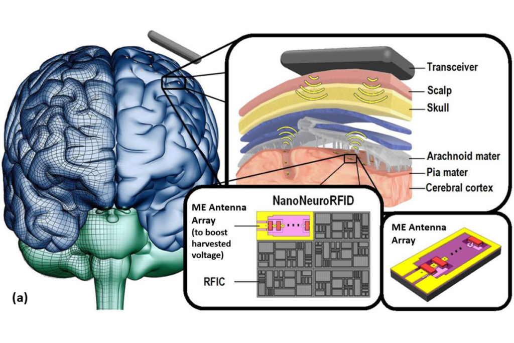
NanoNeuroRFID: A Wireless Implantable Device Based on Magnetoelectric Antennas
Read Description
Electromagnetic-based wireless devices are big in size because of their large antenna, which must be larger than onetenth of the wavelength of the operational frequency. Ultrasound-based wireless devices, in addition to their low data rate, have a massive loss in the skull and need an intermediate electromagnetic transceiver under the skull. Furthermore, almost all state-ofthe- art wireless devices use micro-electrodes for neuronal recording, which are not reliable in long-term monitoring applications because of direct contact between the tissue and metal electrodes. In this paper, we propose a novel wireless and ultra-compact implantable device termed NanoNeuroRFID. At the core of this device there is a Magnetoelectric (ME) antenna array. ME antennas are smart and ultra-miniaturized (<200μm diameter), and can perform multiple tasks: 1) They can harvest electromagnetic energy to power the NanoNeuroRFID system. Their limit of detection for RF magnetic fields is 40pT. 2) They can sense quasi-static neuronal magnetic fields as small as 200pT without a direct contact to the tissue, allowing a long lifetime and reliable neural recording. 3) They can communicate with an external transceiver, and their operational frequency could be 10s to 100s of MHz where tissue loss is small.
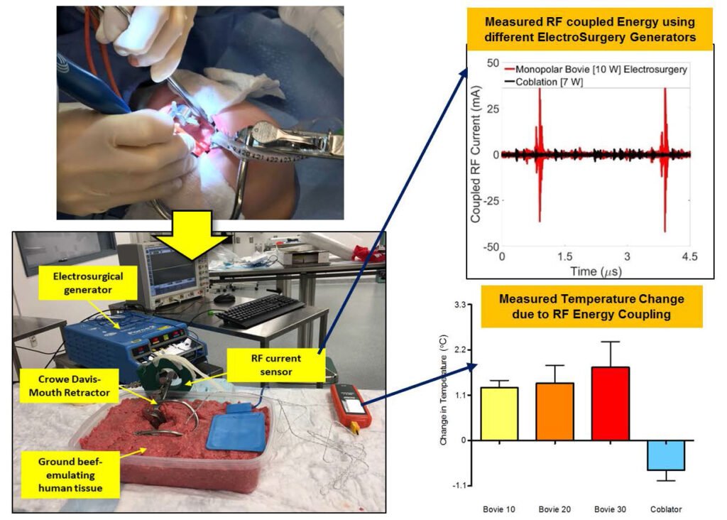
Unintentional RF Energy Transfer During Tonsillectomy: An In Vitro Investigation
Read Description
To validate our hypothesis, in vitro studies are performed in a ground beef phantom with sensors measuring: a) the unwanted current coupled to the mouth retractor, and b) the unwanted temperature rise in the tissues that surround the retractor. The simulated surgery was performed using two separate surgical techniques: monopolar electrosurgery and coblation. Results indicate that unintentional RF energy transfer is indeed a real issue. During electrosurgery, peak-to-peak unwanted currents vary from 80.53 to 181.48 mA for typical power levels ranging from 10W to 30W. Tissue temperature unintentionally increases by 1.3°C and 1.8°C, respectively. Coblation indicates smaller coupling effects, with peak-to-peak currents on the mouth retractor capped at 12.33 mA for a typical 7 W setting. Concurrently, tissue temperature is reduced by 0.73°C, as attributed to the saline solution inherent to coblation. As the first of its kind, this study illuminates possible causes of post-tonsillectomy dysgeusia and intends to trigger future studies. The ultimate goal is safe and complication-free tonsillectomies
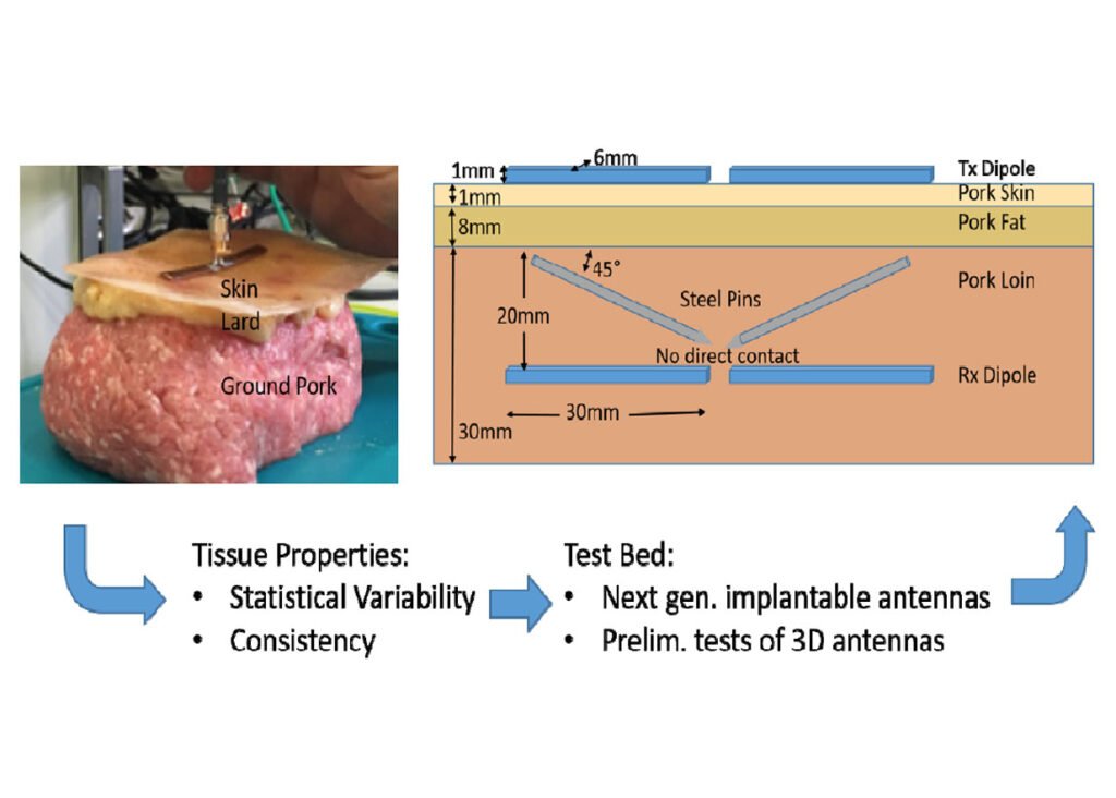
A Layered Pork Model for Subdermal Antenna Tests at 433 MHz
Read Description
Objectives: The objectives of this work are to create a simple biological test bed for subdermal implantable antennas that includes the normal expected variation of the tissues Technology or Method: A layered model using pork products with skin, pork fat or lard, and pork loin or ground pork is used. Results: The ex vivo porcine tissues are similar to in vivo human tissues. Pork fat and loin have more variability than lard and ground pork but are more difficult to imbed subdermal antennas in. This model provides an easy to use platform for testing subdermal antennas in the lab. This test bed was developed for 433 MHz, the ISM band closest to the MedRadio band (402-405 MHz). Initial tests are demonstrated on a two-wire passive system for focusing power within the muscle region.
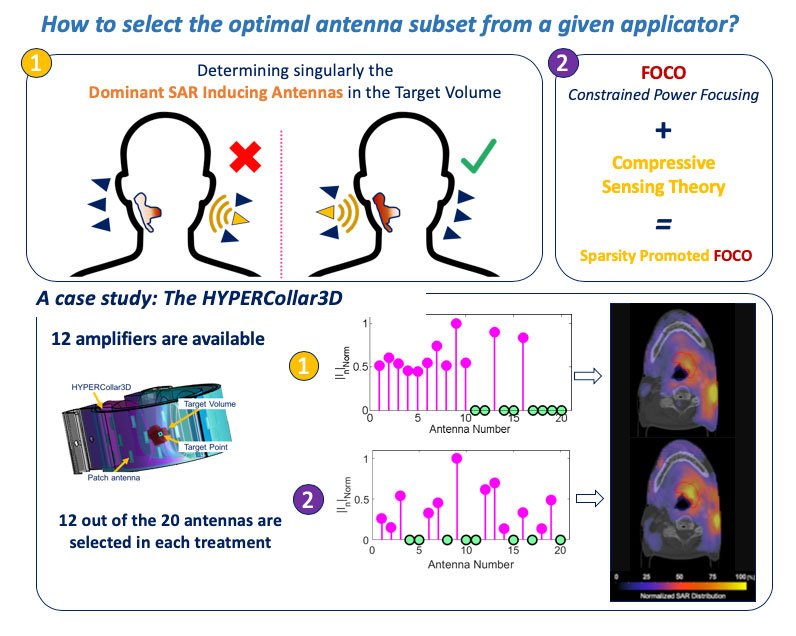
Selecting the Optimal Subset of Antennas in Hyperthermia Treatment Planning
Read Description
Among them, increasing the frequency and the number of radiating elements has shown effective for achieving more conformal heating. However, as each radiating element requires a power amplifier to control it, increasing the number of antennas considerably impacts the overall cost and complexity of the system. Accordingly, a procedure capable of selecting an optimal patient-specific subset of antennas from an oversized phased array applicator (with more antenna elements than available amplifiers) could help improving cost-effectiveness. In this study, we present an original approach which allows improving performance by adaptively selecting the optimal subset of antennas to be activated for a given (redundant) applicator and a given patient. The proposed approach takes inspiration from the compressive sensing theory by embedding the sparsity promotion paradigm into a treatment planning procedure which casts power-deposition as a constrained convex optimization. Performance were demonstrated for the case of head and neck hyperthermia, and benchmarked against the antenna selection procedure presently used in the clinical practice.
RF Injection Network Development for Testing of Active Implantable Medical Devices Exposed to RF Fields in 1.5 T MRI Systems
Read Description
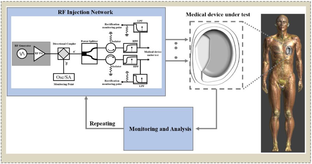
The system was developed to meet the requirements of ISO/TS 10974:2018(E). A directional lumped element coupler, power splitter, an attenuator/isolator, low pass filter and high pass filter were designed and implemented as part of the network. The RF injection network was developed in both a compact version implemented in a single PCB and discrete PCB version for use in different situations. The performance of each designed component was simulated and compared to measurement results. As an application example, a neuromodulation system was tested using the developed RF injection network for conductive emission testing.
Effects of Coaxial-lateral and Coaxial-angular Displacements on Link Efficiency of a Wirelessly Powered Optogenetic Implant: Design, Modeling and Experimental Validation
Read Description
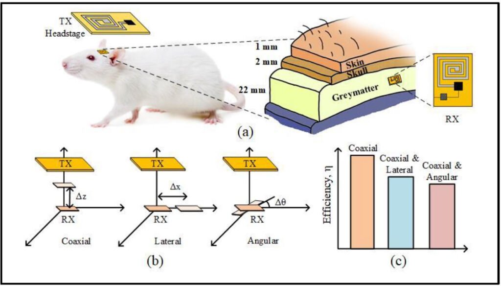
The coupling coefficient deteriorates significantly due to the displacements thus decreasing the overall power transfer efficiency (PTE) of the system. In this paper, we present an analysis and modeling of the effects of various displacements on the efficiency and the overall performance of a miniaturized WPT system designed for an optogenetic implant. To emulate the tissue media inside a human head, skin, skull and gray matter layers are theoretically modeled using dielectric properties and simulation models are developed using the Ansys High Frequency Structure Simulator (HFSS) software. The propagation loss and link efficiency are modeled and simulated as a function of various displacement combinations. To validate the theoretical and simulation models, the WPT system is characterized in various displacement conditions using chicken breast as the media. The measurement results also show a good agreement with the simulation results, thus providing an estimation for the misalignment tolerance range for given specifications. The efficiency performance analysis of the proposed WPT system for various worst-case scenarios also provides a preliminary model for designing a closed-loop wireless power delivery regulation scheme in the future.