Detection of Simulated Brain Strokes Using Microwave Tomography
Read Description
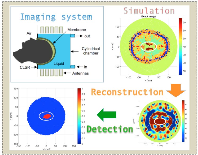
Recent studies in biomedical imaging have shown that strokes produce variations in the complex electric permittivity of brain tissues, which can be detected by means of microwave tomography. Here we present some synthetic results obtained with an experimental microwave tomography-based portable system for the early detection and monitoring of brain strokes. The determination of electric permittivity first requires the solution of a coupled forward-inverse problem. We make use of massive parallel computation from domain decomposition method and regularization techniques for optimization methods. Synthetic data are obtained with electromagnetic simulations corrupted by noise, which have been derived from measurements errors of the experimental imaging system. Results demonstrate the possibility to detect hemorrhagic strokes with microwave systems when applying the proposed reconstruction algorithm with edge preserving regularization.
Low-Cost Ink-Jet Printed RFID Tag Antenna Design for Remote Healthcare Applications
Read Description
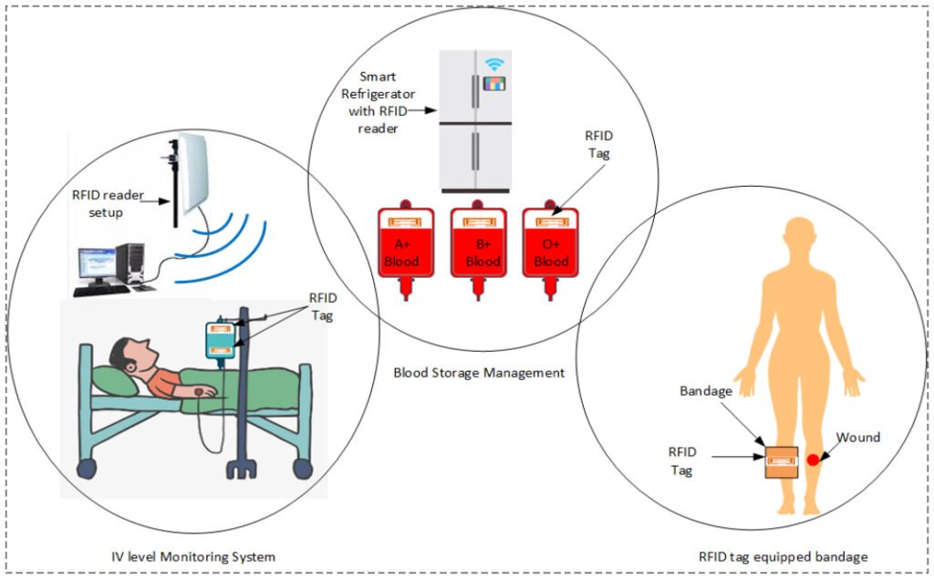
The tag antenna is exploited as a sensor by modifying its equivalent circuit to mitigate the effects of water, blood sample phantom, and the human body. As a result, the proposed RFID antenna features impedance match with Impinj R6 RFID chip from 880 MHz – 937 MHz with compact dimensions of 40 x 14 mm. Moreover, this tag has a read range of 3 m, 2.5 m and 1.5 m on the water bottle, intravenous (IV) solution and blood bag, respectively. However, the read range of RFID tag on an empty water bottle or IV solution bag is 0.5 m. By comparing the read range of tag on empty and solution filled IV bags, the proposed tag is used as a water proximity sensor. Experimental testing of the tag is performed for sensing the level of the IV solution. Also, this tag is tested by mounting on liquid mixture (a mixture of salt and sugar is used as a phantom to mimic the blood) filled plastic bags, which leads to a low-cost solution for blood storage management. Experimental results show a good agreement of proposed tag towards its use in healthcare applications, which leads to better healthcare facilitation regarding cost, time and care.
Quality Control of Microwave Equipment for Tissue Imaging
Read Description
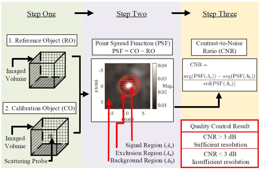
Imaging systems are typically designed with a theoretical resolution limit in mind, which is rarely achieved in practice due to measurement uncertainties, background clutter and system noise. Uncertainties and background clutter are particularly prominent in medical diagnostic imaging. This manuscript proposes a method for data quality assessment of an experimental imaging system that aims at a specific image resolution. It utilizes two measurements, one of a uniform background medium and one of the same medium with a small scattering probe embedded within it. The probe’s size and permittivity reflect the desired application-specific resolution. The method extracts the system point-spread function (PSF) from the two measurements and computes the PSF contrast-to-noise ratio (CNR). A case study is presented, demonstrating the quality control protocol and its ability to identify data sets of inadequate quality and provide an evaluation metric. The protocol also highlights possible sources of error and enables data filtering that increases significantly the reconstructed image quality.
Non-Invasive Wearable RF Device towards Monitoring Brain Atrophy and Lateral Ventricle Enlargement
Read Description
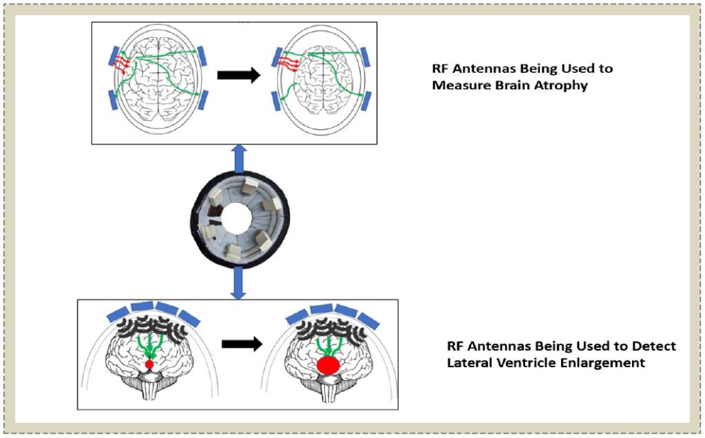
Another effect of Alzheimer’s disease is the enlargement of lateral ventricles in the brain. Currently, MRI and CT scanners can detect and show images of the brain during different stages of Alzheimer’s disease. However, its limited accessibility, high costs, and static structure make it inconvenient for some to use. This paper presents the design and novel application of a wearable device comprising of flexible microwave antennas, with an operating frequency range of 800 MHz to 2.5 GHz, that detects the progression of brain atrophy and lateral ventricle enlargement in patients with Alzheimer’s at the earliest stage possible. The operating principle of the antennas are simulated in near field using CST and the device is experimentally validated using lamb brain samples and samples representing cerebral spinal fluid (CSF). The measured reflection coefficients and transmission coefficients were found to correlate with changes in brain volume and changes in CSF volume successfully, thus giving an indication of the progression of Alzheimer’s disease in a patient.
Smart Textile Integrated Wireless Powered Near Field Communication (NFC) Body Temperature and Sweat Sensing System
Read Description
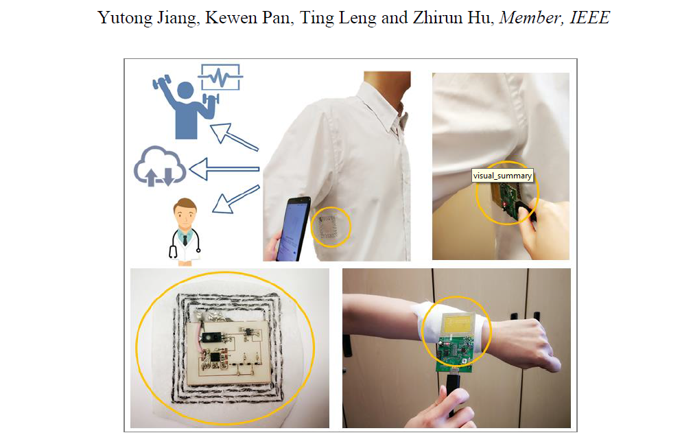
The NFC antenna is seamlessly integrated with closed-body garments, and sensor data can be easily acquired by NFC readers and smart phones in order to achieve real time and wireless monitor of health status in a convenient and non-intrusive way. A Dickson charge pump circuit has been designed and implemented in order to pump up the voltage and ensure a steady voltage supply for the sweat sensor. The maximum read range for accessing sensor data is 6 cm. The on-body measurement accuracy of the temperature sensor and sweat sensor are able to achieve ± 0.14°C and ± 0.2%, respectively. The presented system can provide wearable battery-free ubiquitous wireless connectivity for point-of-care and any time healthcare and wellbeing monitoring.
Focused Microwave Breast Hyperthermia Monitored by Thermoacoustic Imaging: A Computational Feasibility Study Applying Realistic Breast Phantoms
Read Description
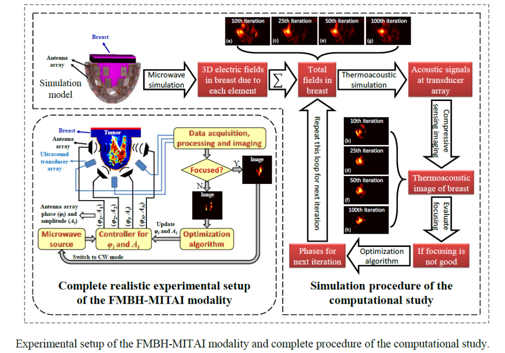
Microwave-induced thermoacoustic imaging (MITAI) is naturally feasible for such power deposition monitoring task. This work conducts a computational study to evaluate feasibility of the novel FMBH-MITAI modality using realistic breast phantoms. Basic configuration and rationale of both FMBH and MITAI are introduced. Com-pressive sensing (CS) technique has to be applied in MITAI for sparse acoustic measurement in the FMBH-MITAI modality. Procedure of the computational study consists of microwave simulation, thermoacoustic numerical simulation, CS imaging, and performing the iterative optimization. Simulated results show that CS based MITAI is able to serve as a reliable monitor-ing mechanism to efficiently guide the iterative optimization toward the best obtainable focusing condition in most of the sim-ulated scenarios. Finally obtained thermoacoustic images agree well with simulated power deposition distribution well. This work offers valuable performance evaluation and is of significant meaning for potential clinical applications of the FMBH-MITAI modality.