Non-Invasive Wearable RF Device towards Monitoring Brain Atrophy and Lateral Ventricle Enlargement
Imran Saied, Tughrul Arslan.
Read Description
Another effect of Alzheimer’s disease is the enlargement of lateral ventricles in the brain. Currently, MRI and CT scanners can detect and show images of the brain during different stages of Alzheimer’s disease. However, its limited accessibility, high costs, and static structure make it inconvenient for some to use. This paper presents the design and novel application of a wearable device comprising of flexible microwave antennas, with an operating frequency range of 800 MHz to 2.5 GHz, that detects the progression of brain atrophy and lateral ventricle enlargement in patients with Alzheimer’s at the earliest stage possible. The operating principle of the antennas are simulated in near field using CST and the device is experimentally validated using lamb brain samples and samples representing cerebral spinal fluid (CSF). The measured reflection coefficients and transmission coefficients were found to correlate with changes in brain volume and changes in CSF volume successfully, thus giving an indication of the progression of Alzheimer’s disease in a patient.
×
Take Out Message
Take-Home Messages
- A wearable device was developed with electromagnetic sensors in order to non-invasively monitor the progress of brain atrophy and lateral ventricle enlargement as a result of Alzheimer’s disease.
- The developed wearable RF device is capable of detecting the progression of brain atrophy and lateral ventricle enlargement successfully.
- The work in this study targets Alzheimer’s disease and aims to develop a non-invasive device for monitoring the progression of the disease in patients.
- The breakthrough in this work is the development of a wearable device that uses RF sensors for detecting changes in the brain as a result of Alzheimer’s disease.
Smart Textile Integrated Wireless Powered Near Field Communication (NFC) Body Temperature and Sweat Sensing System
Yutong Jiang, Kewen Pan, Ting Leng, Zhirun Hu.
Read Description
The NFC antenna is seamlessly integrated with closed-body garments, and sensor data can be easily acquired by NFC readers and smart phones in order to achieve real time and wireless monitor of health status in a convenient and non-intrusive way. A Dickson charge pump circuit has been designed and implemented in order to pump up the voltage and ensure a steady voltage supply for the sweat sensor. The maximum read range for accessing sensor data is 6 cm. The on-body measurement accuracy of the temperature sensor and sweat sensor are able to achieve ± 0.14°C and ± 0.2%, respectively. The presented system can provide wearable battery-free ubiquitous wireless connectivity for point-of-care and any time healthcare and wellbeing monitoring.
×
Take Out Message
Take-Home Messages
- In this manuscript, body temperature and sweat sensors are integrated with a textile NFC antenna, which eliminates the need for external batteries and realizes real-time wireless monitoring.
- This paper has presented design, fabrication implementation, measurements and real-life applications of smart textile NFC antennas and a battery-free wireless NFC body temperature and sweat sensing device, aiming for truly ubiquitous wireless health and wellbeing monitoring.
- The proposed device targets at body temperature and sweat loss monitoring for daily healthcare, systemic hyperthermia from fever, sweating symptoms caused by various kinds of infection, inflammation and trauma and wound healing monitoring.
- Different from conventional battery enabled and wire connected sensors, the significance of this work is by applying textile NFC as a communication interface as well as a wireless power harvester, battery-free real-time body temperature and sweat monitoring has been realized simultaneously.
- Apart from the device itself, an App has also been developed on Android system for the sensor data to be accessed by smart phones.
Focused Microwave Breast Hyperthermia Monitored by Thermoacoustic Imaging: A Computational Feasibility Study Applying Realistic Breast Phantoms
Lifan Xu, Xiong Wang.
Read Description
Microwave-induced thermoacoustic imaging (MITAI) is naturally feasible for such power deposition monitoring task. This work conducts a computational study to evaluate feasibility of the novel FMBH-MITAI modality using realistic breast phantoms. Basic configuration and rationale of both FMBH and MITAI are introduced. Com-pressive sensing (CS) technique has to be applied in MITAI for sparse acoustic measurement in the FMBH-MITAI modality. Procedure of the computational study consists of microwave simulation, thermoacoustic numerical simulation, CS imaging, and performing the iterative optimization. Simulated results show that CS based MITAI is able to serve as a reliable monitor-ing mechanism to efficiently guide the iterative optimization toward the best obtainable focusing condition in most of the sim-ulated scenarios. Finally obtained thermoacoustic images agree well with simulated power deposition distribution well. This work offers valuable performance evaluation and is of significant meaning for potential clinical applications of the FMBH-MITAI modality.
×
Take Out Message
Take-Home Messages
- Microwave-induced thermoacoustic imaging (MITAI) is applied to monitor microwave power deposition distribution in human breast during the process of focused microwave breast hyperthermia (FMBH).
- Compressive sensing (CS) based MITAI technique is able to provide reliable power deposition monitoring for the iterative focusing process of the FMBH approach under the condition that the obtainable focusing is good enough.
- Focused microwave breast hyperthermia (FMBH) for treating breast tumors noninvasively.
- This work presents the first systematic computational study for assessing performance and robustness of the MITAI monitored FMBH modality, referred to as FMBH-MITAI modality, utilizing realistic human breast phantoms with different densities and tumor locations.
Microwave Dielectric Sensing of Free-Flowing, Single, Living Cells in Aqueous Suspension
Clare Watts, Stephen Hanham, James Armstrong, Munir Ahmad, Molly Stevens, Norbert Klein.
Read Description
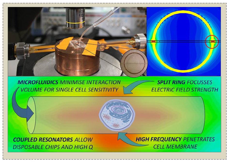
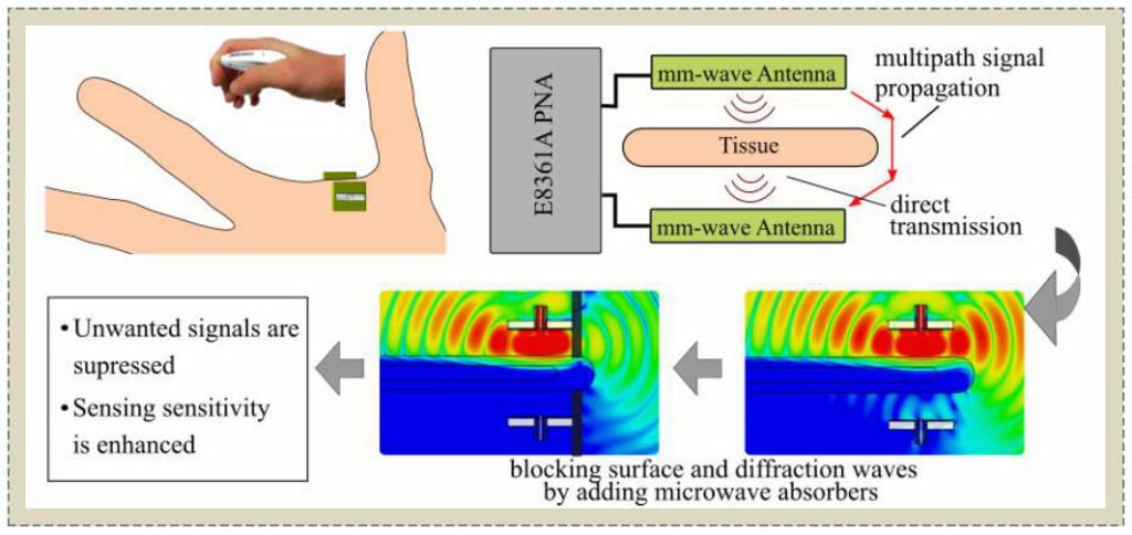
Inductive coupling between the two resonators enabled separation of microfluidic chips from RF connectors and allowed for time-resolved continuous-wave measurements on flowing single cells via the coaxial ports of a dielectric-loaded microwave cavity. Analysis via an equivalent circuit model showed that the novel resonator assembly maintained the permittivity-dependent sensitivity of a split ring resonator while operating at quality factors >1000 with lossy aqueous media (typically ~1900). Using a microfluidic channel with a 300 x 300 μm cross section, at a water-loaded resonant amplitude of ~-22 dB at 0 dBm input power level, shifts in amplitude due to individual cells passing through the sensing region of up to -0.0015 dB were observed. Correlations between averaged amplitude shifts and cell size as well as material properties demonstrate the diagnostic potential of this technique.
×
Take Out Message
Take-Home Messages
- An inductively-coupled resonator assembly is shown to allow the detection of significantly sub-wavelength diameter biological cells by combining the strong field confinement provided by a split ring resonator with the high quality factor resonance of a dielectric resonator.
- Measurements of single, free-flowing cells in a natural aqueous environment at ~10 GHz have been carried out using a coupled resonator sensor, without the need for trapping, immobilizing, culturing or fixing cells in high-field areas.
- The coupled resonator approach proposed in this work shows potential as a method of discrimination of cells based on hydration levels, which in other works has been linked to carcinogenesis, as well as cancer aggressiveness grade; therefore the sensor described herein may represent an alternative method of cancer diagnosis or disease progression monitoring via non-invasive liquid biopsies.
- In this paper, measurements of living, free-flowing, single cells in aqueous buffer solution, at a frequency sensitive to cell water content, have been made.
- The inductive coupling employed in this sensor allows for physical separation of the sensing elements from microwave electronics, allowing for cheap, disposable chips to be used with biological fluids.
Photovoltaic Power Harvesting Technologies in Biomedical Implantable Devices Considering the Optimal Location
Jinwei Zhao, Rami Ghannam, Man Kay Law, Muhammad Ali Imran, Hadi Heidari.
Read Description
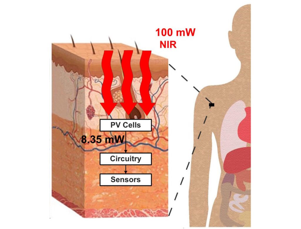
For example, current energy harvesters rely on piezoelectricity, thermoelectricity and solar electricity to drive the implantable device. However, the majority of these energy harvesting techniques suffer from a variety of limitations such as low power output, large size or poor efficiency. Due to their high efficiency, we focus our attention on solar photovoltaic cells. We demonstrate the tissue absorption losses severely influence their performance. We predict the performance of these cells using simulation through the verified experimental data. Our results show that our model can obtain 17.20% efficiency and 0.675 V open-circuit voltage in one sun condition. In addition, our device can also harvest up to 15 mW/ cm2in dermis and 11.84 mW/ cm2in hypodermis by using 100 mW/ cm2light source at 800 nm and 850 nm, respectively. We propose implanting our device in hypodermis to obtain a stable power output.
×
Take Out Message
Take-Home Messages
- This is first time to model the performance of implantable PV cells in different layers of tissue. We demonstrate how the electrical characteristics are influenced by the implanting location of the device.
- A PV cell implanted in the dermis layer can harvest the greatest amount of power.
- We propose implanting our energy harvesting PV cells in the hypodermis layer.
- Our proposed PV device harvests enough energy to supply power for low-cost implants such as cardiac pacemakers, retinal implants or biomedical sensors.
- PV cells implanted in the adipose layer can harvest nearly 11.84 mW using an 850 nm light source.
Comparison of Different Assessment Quantities to Evaluate Lead Electromagnetic Model for Radio Frequency Energy-Induced Heating
Mikhail Kozlov ; Wolfgang Kainz.
Read Description
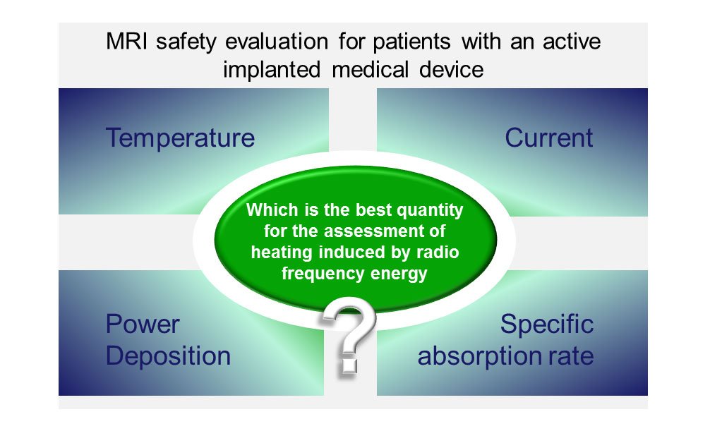
The set included 32 single electrode leads with straight and helical wires. The LEMs were obtained and validated with 3D electromagnetic and thermal co-simulations at 128 MHz. A shift of the position of temperature or SAR sensor from the electrode tip to the electrode pedestal resulted in a decrease of the linear regression coefficient of determination for the LEM calibration factors. Behavior of different assessment quantities in terms of sensitivity of sensor location significantly varied between leads with the helical and straight wires. In conclusion, utilization of analysis made for generic leads with straight wire can be significantly misleading for the prediction of results for leads with helical wires.
×
Take Out Message
Take-Home Messages
- Four quantities, namely 1) the net dissipated power around an electrode of an active implantable medical device (AIMD), 2) the net temperature increase, 3) the current flowing from the lead into the electrode, and 4) the net specific absorption rate (SAR) increase, were numerically compared for a set of leads with straight and helical wires to evaluate the lead electromagnetic model (LEM) with respect to radio frequency energy-induced heating.
- The most suitable sensor locations were positions from the electrode tip along the first half of the electrode axial axis. A temperature sensor was essential if the net electrode temperature increase required evaluation.
- The targeted medical application is an evaluation of heating induced by radio frequency energy that appears in human tissue near an AIMD during magnetic resonance imaging.
- For leads with helical wire, our results indicate that 1) to achieve a good validation of the transfer function, that is, the linear regression coefficient of determination R2 to be close to 1, the temperature sensor results must be obtained as fast as possible, but 2) the total transient time must be longer than 360 seconds for evaluation of the LEM calibration factor.
- Utilization of result analysis made for generic leads with straight wire can be significantly misleading for predicting results for leads with helical wires.