Realization and Experimental Assessment of Baseball-Bat Microwave Antenna for Low Power Cancer Ablation
Read Description
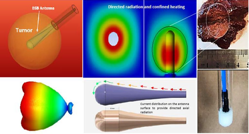
Development of baseball-bat shaped (BSB) antenna has been studied using electromagnetic and thermal simulations and evaluated experimentally. Numerical simulations showed less than -10-dB reflection stability is attained for more than 20 GHz. Electromagnetic simulation showed that highly directed end-fire radiation achieves confined power deposition within targeted model and yields in higher SAR attained. Nearly spherical ablated lesions are achieved with no healthy tissues being destroyed in the backward direction. Proposed antenna was fabricated and tested in ex-vivo bovine liver sample and egg-white solution. Good agreement between simulated and measured results where confined ablated lesions attained at only 1W were comparable to that obtained at much higher power ranges (20-60W). Efficacy of BSB antenna to efficiently radiate in different dielectric mediums is noticeably attained. The proposed antenna model may help improving the precision of microwave ablation associated with commonly broadside radiators previously used in literature and provide homogenous SAR and confined heating to overcome the limitations found in treating spherical tumors with heterogeneous properties using much high power with narrow-band feature.
An invivo-mimickingIn vitrotestbed for brain-computer interfaces
Read Description
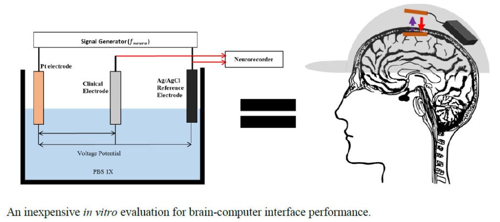
As such, optimizing and testing via function generators and resistors overestimates BCI sensitivity by at least 250 times and 2 times, respectively. To more closely replicate a clinical recording environment and accurately assess BCI performance, we utilize a function generator to mimic neural signals sent to a human-body-mimicking electrolyte fluid via a Pt electrode. These signals are then recorded via a neural electrode connected to the BCI under test. To validate the proposed method, we compare sensitivity of a previously reported fully passive BCI via the three methods. The presented results demonstrate that recording via the proposed clinically accurate setup when the BCI has been optimized with one of the two other test methods gives the lowest sensitivity in the frequency domain and worst signal stability in the time domain. Overestimating BCI sensitivity during in vitro testing results in the potential inability to record desired neural signals in vivo. The proposed method provides a means by which to accurately assess BCI performance, therefore reducing errors during animal trials and saving time, money, and lives.
Influence of low frequency Near-Field Sources Position on the Assessment of Children Exposure Variability using Stochastic Dosimetry
Read Description
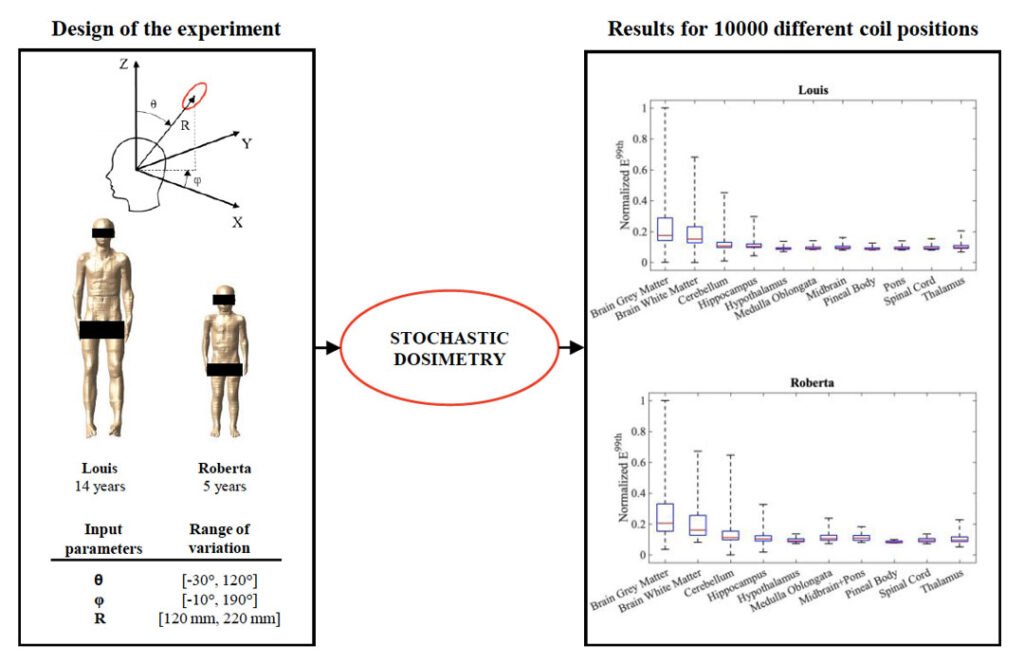
In details, in this paper the exposure of two child models to a hairdryer model was evaluated. Following the ICNIRP guidelines, the electric field amplitudes induced in specific tissues composing the central nervous system and the peripheral nervous system were analyzed. The analysis of the results permitted to highlight a high exposure variability depending on the near-field source position and to individuate the regions where the source could cause the highest levels of exposure, not limiting the analysis only to some worst-case exposure scenarios.
Cardiac Influence of Repetitive Transcranial Magnetic Stimulation in Small Animals
Read Description
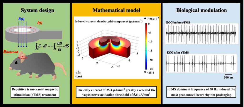
However, the potential adverse effects of rTMS on heart rhythm have not been extensively investigated. This study aims to develop an optimized design of rTMS system to evaluate the potential adverse effects of rTMS on mouse heart rhythm via vagus nerve modulation for pre-clinical application. The rTMS-induced electric field in the vagus nerve of brain produced by the strong rate of current change of 1.04×10⁸ A/s in a stimulating coil, which was directly determined by circuit design in charging voltage of the capacitor bank and inductance value of a stimulating coil. A finite element method (FEM) mathematical simulation indicated that the maximum eddy current was 25.4μA/mm2, which was greatly exceeded the vagus nerve activation threshold of 5.6μA/mm2. The animal experiment results also verify that the induced electric field activates the RR-interval prolonging effect might be attributed to vagus nerve stimulation (VNS) from rTMS, and the most pronounced heart rhythm prolonging effect at 20Hz magnetic field treatment, causing the average heart rate decreased to 58.65% of that before rTMS in 10 mice. In conclusion, above-threshold rTMS at 20 Hz could produce maximum adverse effect on heart rhythm through direct vagus nerve activation for pre-clinical applications such as safety screening.
Robustness of Time-Multiplexed Hyperthermia to Temperature Dependent Thermal Tissue Properties
Read Description
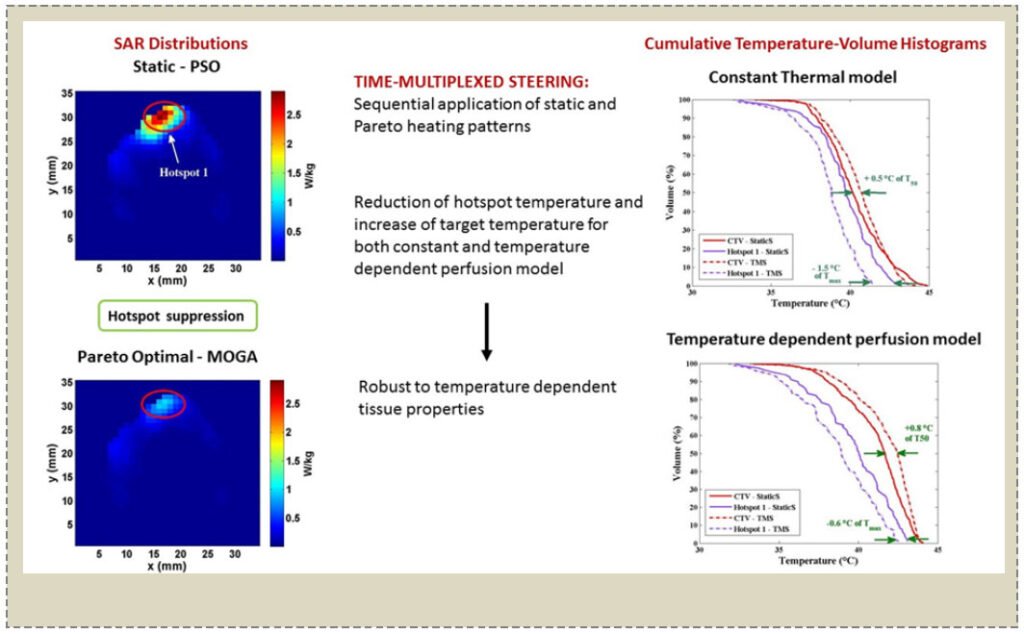
The principal need in hyperthermia treatment is to optimally focus the heating into the target while minimising heating in the surrounding healthy tissue. Patient-specific treatment planning is done to optimize the specific absorption rate and the resulting temperature distribution. Uncertainties associated with the thermal model used for temperature simulations represent an important challenge. Our previous work has demonstrated that the occurrence of hotspots can be reduced and target heating enhanced using time-multiplexed steering procedures. In this paper, the robustness of time-multiplexed hyperthermia against temperature dependent thermal tissue properties is investigated. Temperature simulations are used to predict the time-dependent heating achieved by multiple antenna phase and amplitude configurations that are generated by a multi-objective genetic algorithm and applied sequentially. The proposed strategy is compared with the heating obtained using one single heating setting obtained by particle swarm optimization as typically used in clinical hyperthermia. Thermal performance of the static and time-multiplexed methods are assessed by applying two thermal models, one that uses constant properties of blood perfusion and thermal conductivity of tumor, muscle and fat, and a second one that uses temperature dependent perfusion values. This study shows that time-multiplexed hyperthermia enhances target heating and limits the hotspot appearance regardless of the thermal model used in thermal simulations.
Detection and Monitoring of Osteoporosis in a Rat Model by Thermoacoustic Tomography
Read Description
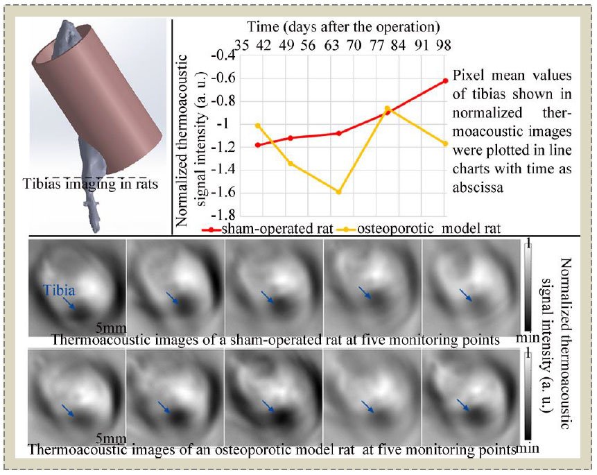
Therefore, here we propose to use TAT to detect osteoporosis and to monitor the formation of osteoporosis over a long period. Technology or Method: This study used the bilateral ovariectomy to obtain an osteoporotic rat model (n=4) along with a sham control. During the 100 days after the operation, the right tibia of each rat was in vivo thermoacoustically imaged at 5 time points. After the last TAT imaging, micro computed tomography (Micro-CT) was performed on each rat to validate the TAT findings. Visual observation and semi-quantitative methods were used to analyze the thermoacoustic data. Results: During the monitoring period, the thermoacoustic signal intensity of the tibia of the sham-operated rat continued to increase, while the thermoacoustic signal intensities of the tibia of osteoporotic model rats showed fluctuations. The TAT findings were verified by Micro-CT. Conclusions: Osteoporosis can be clearly detected by TAT. Significant differences in thermoacoustic signal intensities between normal bone growth and osteoporotic bone formation are observed. Clinical or Biological Impact: This study provides initial facts that TAT may become a new tool for noninvasive detection and monitoring of osteoporosis.