Cardiac Influence of Repetitive Transcranial Magnetic Stimulation in Small Animals
Ting-Wei Wang, Yen-Ling Sung, Shien-Fong Lin.
Read Description
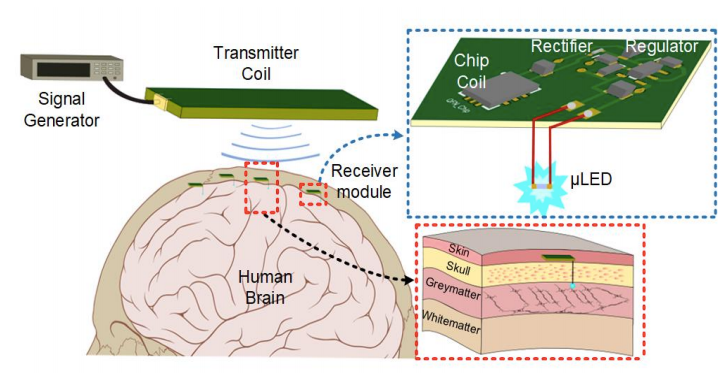
However, the potential adverse effects of rTMS on heart rhythm have not been extensively investigated. This study aims to develop an optimized design of rTMS system to evaluate the potential adverse effects of rTMS on mouse heart rhythm via vagus nerve modulation for pre-clinical application. The rTMS-induced electric field in the vagus nerve of brain produced by the strong rate of current change of 1.04×10⁸ A/s in a stimulating coil, which was directly determined by circuit design in charging voltage of the capacitor bank and inductance value of a stimulating coil. A finite element method (FEM) mathematical simulation indicated that the maximum eddy current was 25.4μA/mm2, which was greatly exceeded the vagus nerve activation threshold of 5.6μA/mm2. The animal experiment results also verify that the induced electric field activates the RR-interval prolonging effect might be attributed to vagus nerve stimulation (VNS) from rTMS, and the most pronounced heart rhythm prolonging effect at 20Hz magnetic field treatment, causing the average heart rate decreased to 58.65% of that before rTMS in 10 mice. In conclusion, above-threshold rTMS at 20 Hz could produce maximum adverse effect on heart rhythm through direct vagus nerve activation for pre-clinical applications such as safety screening.
×
Take Out Message
Take-Home Messages
- Theoretically and experimentally evaluate important parameters such as current and stimulating frequencies in repetitive transcranial magnetic stimulation (rTMS) that could modulate heart rhythm.
- Our system generated an eddy current of 25.4 μA/mm2 in the mouse brain regions and produced the maximum heart rhythm modulating effect at 20 Hz.
- The combined modeling and experimental approach is applicable to explore the potential adverse effects of exogenous electromagnetic fields on heart rhythm.
- Our study provides novel insights into the mechanism of heart rhythm modulation through rTMS and demonstrates the quantitative and morphological aspects of ECG alteration in such outcome.
- The rTMS dominant frequency of 20 Hz induced the most pronounced heart rhythm prolongation, causing the heart rate to decrease by 58.65 % compared to that before rTMS.
Robustness of Time-Multiplexed Hyperthermia to Temperature Dependent Thermal Tissue Properties
Grazia Cappiello, Margarethus Paulides, Tomas Drizdal, Declan Oloughlin, Martin Ohalloran, Martin Glavin, Gerard Van Rhoon, Edward Jones.
Read Description
The principal need in hyperthermia treatment is to optimally focus the heating into the target while minimising heating in the surrounding healthy tissue. Patient-specific treatment planning is done to optimize the specific absorption rate and the resulting temperature distribution. Uncertainties associated with the thermal model used for temperature simulations represent an important challenge. Our previous work has demonstrated that the occurrence of hotspots can be reduced and target heating enhanced using time-multiplexed steering procedures. In this paper, the robustness of time-multiplexed hyperthermia against temperature dependent thermal tissue properties is investigated. Temperature simulations are used to predict the time-dependent heating achieved by multiple antenna phase and amplitude configurations that are generated by a multi-objective genetic algorithm and applied sequentially. The proposed strategy is compared with the heating obtained using one single heating setting obtained by particle swarm optimization as typically used in clinical hyperthermia. Thermal performance of the static and time-multiplexed methods are assessed by applying two thermal models, one that uses constant properties of blood perfusion and thermal conductivity of tumor, muscle and fat, and a second one that uses temperature dependent perfusion values. This study shows that time-multiplexed hyperthermia enhances target heating and limits the hotspot appearance regardless of the thermal model used in thermal simulations.
×
Take Out Message
Take-Home Messages
- A time-multiplexed hyperthermia treatment planning technique aiming at focusing the tumor heating while protecting the healthy tissue is evaluated with temperature-dependent tissue properties.
- The time-multiplexed hyperthermia via MOGA optimization can successfully intensify the heating into the target region while suppressing pre-defined hotspots when either constant thermal properties or temperature dependent tissue properties are assumed.
- The targeted medical application is hyperthermia treatment planning in order to maximize heating in the tumor while minimizing heating in surrounding tissues.
- This work demonstrates the robustness of the time-multiplexed hyperthermia approach to the variation of tissue properties due to temperature increases and ensures the clinical benefit of the method.
- This work demonstrates that time-multiplexed hyperthermia is effective, regardless of the thermal model used.
Detection and Monitoring of Osteoporosis in a Rat Model by Thermoacoustic Tomography
Zihui Chi, Xiao Liang, Xue Wang, Lin Huang, Huabei Jiang.
Read Description
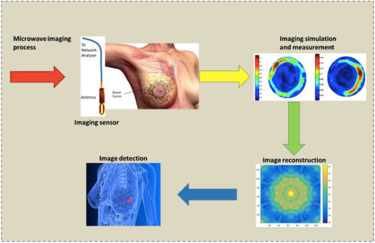
Therefore, here we propose to use TAT to detect osteoporosis and to monitor the formation of osteoporosis over a long period. Technology or Method: This study used the bilateral ovariectomy to obtain an osteoporotic rat model (n=4) along with a sham control. During the 100 days after the operation, the right tibia of each rat was in vivo thermoacoustically imaged at 5 time points. After the last TAT imaging, micro computed tomography (Micro-CT) was performed on each rat to validate the TAT findings. Visual observation and semi-quantitative methods were used to analyze the thermoacoustic data. Results: During the monitoring period, the thermoacoustic signal intensity of the tibia of the sham-operated rat continued to increase, while the thermoacoustic signal intensities of the tibia of osteoporotic model rats showed fluctuations. The TAT findings were verified by Micro-CT. Conclusions: Osteoporosis can be clearly detected by TAT. Significant differences in thermoacoustic signal intensities between normal bone growth and osteoporotic bone formation are observed. Clinical or Biological Impact: This study provides initial facts that TAT may become a new tool for noninvasive detection and monitoring of osteoporosis.
×
Take Out Message
Take-Home Messages
- Thermoacoustic tomography (TAT) is applied to detect and monitor the formation of osteoporosis for the first time, and to further explore the physiological mechanism of osteoporosis formation from the perspective of tissue dielectric properties.
- Significant differences in thermoacoustic signal intensities between normal bone growth and osteoporotic bone formation are observed, suggesting that TAT has the potential to detect and monitor osteoporosis.
- TAT can provide useful information for diagnosis of osteoporosis, prediction of fracture risk, and monitoring of disease progression.
- This study represents the first for TAT to in vivo image osteoporosis and provides initial facts that TAT may become a new tool for noninvasive detection and monitoring of osteoporosis.
- This work is an exploratory experimental study of TAT for imaging osteoporosis, using micro-CT to validate the TAT findings.
Comparison of Radio Frequency Current and Microwave Energy for Transcatheter Renal Denervation
Aditya Rakhmadi, Kazuyuki Saito, Shohei Matsuhara, Tomoyuki Tajima, Nobuyoshi Takeshita.
Read Description
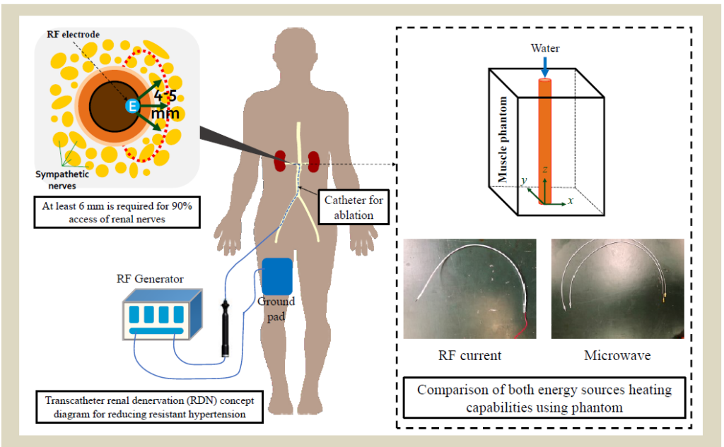
We present the feasibility of using microwave energy as a different energy source for RDN ablation treatment, by comparing to RF current energy source. In this paper, we designed a coaxial-slot antenna for microwave (2.45 GHz) and an electrode for RF current (500 kHz), with phantoms that match human muscle dielectric properties at each respective working frequency. Both phantoms thermal properties and material density were measured, and then used in simulation. A total of 10 ablation experiments were conducted to validate the numerical simulation results. Each temperature points of the experiments were measured at 1 mm interval between thermometer probes. The results of the experiment temperature distributions agree well with the simulation results, RF achieved 5-6 mm while microwave achieved 7-8 mm. Additionally, numerical calculations with different input power were conducted to understand the temperature characteristics of both energy source. Microwave energy offers higher input power for a more profound and broader ablation area as opposed to RF current.
×
Take Out Message
Take-Home Messages
- Utilizing microwave energy, a deeper, four-quadrant ablation area for transcatheter renal denervation (RDN) can be realized without causing complicated vascular lesions as opposed to widely used radio frequency current devices.
- An ablation depth of 8 mm or more with a lower maximum temperature of 65 ºC was achieved by using microwave energy as opposed to radio frequency currents 4-5 mm depth and 87 ºC maximum temperature, shown by numerical calculation and heating experiments.
- The potential application of this microwave technology is the RDN, aimed at reducing resistant hypertension.
- Main contribution of this paper is by comparing microwave energy to radio frequency current based devices in RDN treatment, both in numerical calculations and heating experiments, it is possible to overcome performance shortcomings of widely used devices right now.
- In addition, muscle phantoms at 500 kHz and 2.45 GHz respectively, was produced and both phantoms properties were measured to match parameters used in the numerical calculations and the heating experiments.
Modelling of the temperature changes induced by transcutaneous spinal direct current stimulation (tsDCS)
Serena Fiocchi, Emma Chiaramello, Alberto Priori, Paolo Ravazzani, Marta Parazzini.
Read Description
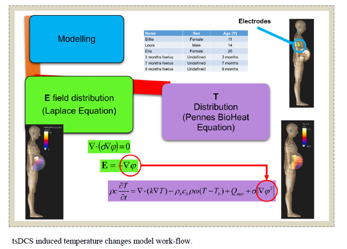
In this study, the temperature increase induced by a 3 mA stimulation in the tissue target (i.e. the spinal cord), in its surrounding tissues and in other tissues possibly more vulnerable to temperature increase were assessed through a computational approach. That solves Laplace equation and BioHeat equation for electric field and temperature distribution, respectively, on six whole body high resolution anatomical models, including three models of pregnant women at different gestational ages. Results show that temperature increase distribution in those targets is guided by a complex interaction between different mechanism in which electric stimulation plays a secondary role, in particular when blood perfusion is active. The very low heating assessed (below 1.5 m°C) is not consistent with the hypothesis that the induced temperature increase would critically activate metabolic changes in the target tissues here considered or would contribute to the few side effect that the applications of spinal direct current stimulation protocols have shown.
×
Take Out Message
Take-Home Messages
- This study deals with safety thermal issues related to the application of spinal tDCS.
- Combined resolution of Laplace and bio-heat equation allows to assess temperature increase due to electric stimulation induced by spinal tDCS.
- This is the first study addressing the tissues temperature changes induced by tsDCS.
- The very low heating induced by tDCS is not likely to activate metabolic changes in the target tissues here considered or to contribute to the few side effect that the applications of spinal tDCS protocols have shown.
- Findings of this work respond to the need of evaluating the safety of the spinal tDCS application on different subjects (young male, female, and pregnant women).
Field Focusing for Implanted Medical Devices
Hossein Mehrpour Bernety, Huanan Zhang, David Schurig, Cynthia M. Furse.
Read Description
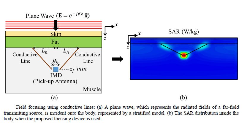
The small gap between the tips of the lines provides constructive coupling and augments the focused field. Compared to the case where no lines are used, the focused power is 8 times and 16 times larger if one line and two lines are utilized, respectively. The proposed design can be modified to focus fields with arbitrary polarizations at different depths inside the body. We propose a method to create this focusing structure by extrusion inside the body using heat-activated polymer-based conductors.
×
Take Out Message
Take-Home Messages
- We present a novel method of electromagnetic field focusing applicable to 3D implantable devices.
- Compared to the case of no focusing device, the achieved focused power is 8 and 16 times greater when the proposed designs are used.
- It could improve telemetry or wireless power transfer for future miniaturized implantable medical devices (IMDs).
- The paper presents a novel field focusing method, which is simple and biocompatible applicable to 3D implantable devices.
- The proposed design can be modified to focus fields with arbitrary polarizations at different depths inside human tissue.