Non-Contact Human Gait Analysis and Limb Joint Tracking Using Doppler Radar
Farhan Quaiyum, Nghia Tran, Jean E. Piou, Ozlem Kilic, Aly Fathy.
Read Description
Here, we investigate the feasibility of extending the 1-D block processing algorithm to distinctly track specific limb joints and discuss the advantages and limitations of the technique for CW radar. To establish a repeatable reference data, we run a full wave EM analysis on customized Boulic human model emulating specific video recorded body motions. Results based on measured data are in agreement with simulated ones for simple motions like swinging one hand or one leg only. The proposed technique is also successful in extracting lower body parts while the whole body is in motion, however it is still hard to clearly extract upper body parts like swinging hands.
×
Take Out Message
Take-Home Messages
- The developed EM model for human motions creates repeatable and realistic reference data which when coupled with the signal processing technique can be useful for obtaining a full understanding of human limb joint motion analysis.
- The proposed method is successful in extracting different limb joint trajectories from a complex human motion but with some limitations such as difficulty in tracking hands for a walking subject and reliance on reference data to identify the desired motion details.
- The targeted applications of this work are treating patients with joint problems, athlete performance analysis, motion classification, and so on.
- The significance of this work is the development of a limb joint tracking technique suitable for use with low cost and simple Doppler radar in a typical non-controlled environment.
- A by-product of this work is the use of a portable and flexible software-defined transceiver system as the Doppler radar utilized in the experiments.
Mesoporous titania-coated biosensor and FEM model design for highly sensitive detection of low molecular weight targets
Ollivier Tamarin, Hamida Hallil Abbas, Wassim Ouelhazi, Maxence Rube,Jean Luc Lachaud, Vincent Raimbault, Cedric Boissiere, Marie Paule Bonnet,Dominique Rebiere & Corinne Dejous.
Read Description
The strategy of using such Love wave devices, with 3D porous layers, offering further easy functionalization, aims not only to increase the amount of targets caught on the sensor surface, but also to enhance the detection mechanism by a higher perturbation of the Love wave acoustic energy which could be trapped inside the 3D sensitive layer. First, as a proof of concept, experimental devices with a 3D titania mesoporous layer were realized, and they have shown a good agreement with simulated results. Furthermore, experimental test with several Newtonian liquids are investigated, in a range of viscosities from 1 to 7 cP, typical of those concerned by our biochemical applications. The sensitivity with a 300 nm thick porous sensing layer was 10 times that of the bare device, with interesting dynamical issues to be further studied, giving rise to the great potentialities for biological detection of low weight biochemical targets.
×
Take Out Message
Take-Home Messages
- We present an easy-to-use, portable, autonomous RF electronic readout circuit as a first proof-of-concept for a passive biosensor network platform that integrates passive acoustic wave biosensors and their remote interrogation.
- The acoustic device can detect low-weight biochemical targets in low-volume samples and represents an important step toward a biosensor network platform for cancer diagnosis and monitoring of environmental health.
- The association of a Love wave device with functionalized porous matrices could improve the performance of the sensor in biological media for highly sensitive detection of low weight molecular targets while facilitating the development of an easily regenerable system.
- The main innovation is related to the modelling of the device and simulation using the Finite Element Method (FEM), which is a good way to take into account the physical properties of porous 3D-layers which would also make it possible to design very sensitive layers adapted to the detection targets, such as cancer bio-markers and toxins.
- This novel approach has potential applications low molecular weight biochemical detection for early cancer diagnosis and environmental monitoring, among others.
Combined Effect of 60 Hz Magnetic Fields and Anticancer Drugs on Human Hepatoma HepG2 Cells
Makiko Kakikawa, Tetsuya Maeda, Sotoshi Yamada.
Read Description
However, since the prokaryotic bacterial cells, E.coli, differ from human cells which are eukaryotes and multicellular organisms in many ways, it was not clear whether the magnetic fields affect a potency of anticancer drug against human cancer cells. In this study, we designed and produced magnetic fields exposure device for human cancer cells in culture, and investigated whether 60 Hz, 50 mT magnetic fields affect the potency of anticancer drugs against human hepatoma HepG2 cells. The results of experiments with an anticancer drug, cisplatin indicated that the quantity of viable HepG2 cells become decreased significantly by the combination of cisplatin and magnetic fields as compared to that by cisplatin alone. This suggested that 60 Hz, 50 mT magnetic fields increase the cytocidal activity of cisplatin to human hepatoma cells. The efficiency of the anticancer drugs, mitomycin C and doxorubicin against HepG2 cells was also increased significantly by exposure to magnetic fields, although the time associated with the greatest enhancement of the drugs potency achieved by magnetic fields differed among drugs. These results suggest that 60 Hz, 50 mT magnetic fields strengthen the effect of anticancer drugs on human cancer HepG2 cells.
×
Take Out Message
Take-Home Messages
- The magnetic fields exposure device was newly designed and produced for generating magnetic fields in a CO2-incubator culturing of human cells.
- The results suggested that 60 Hz, 50 mT magnetic fields enhance the efficacy of anticancer drug to human cancer cells.
- Three types of anticancer drugs used in this study are affected differently by the magnetic fields. The magnetic fields increase the effects for all the drugs, by which the numbers of viable cells are decreased by 40% than those with only the drugs alones.
- If our finding can be applied clinically, magnetic fields exposure to cancer site might allow for an effective target chemotherapy, reducing dosage and suppressing side effects.
Application of Two-Dimensional Discrete Dipole Approximation in Simulating Electric Field of a Microwave Breast Imaging System
Samar Hosseinzadegan, Andreas Fhager, Mikael Persson, Paul Meaney.
Read Description
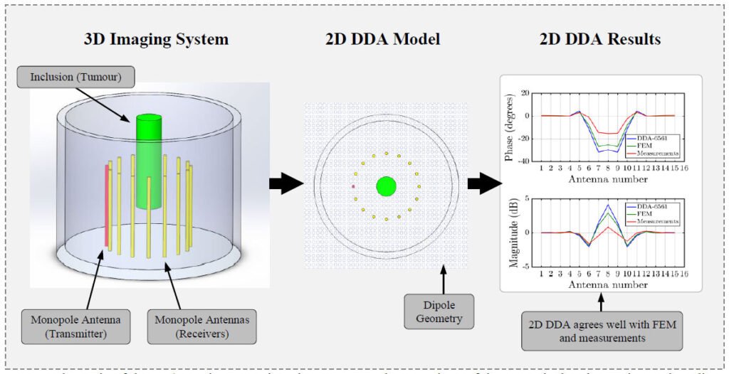
We have modeled the field distributions in COMSOL Multiphysics as baseline results to benchmark the DDA simulations. We have also investigated the adequate sampling size and the effect of inclusion size and property contrast on solution accuracy. In this way, we can utilize the 2D DDA as an alternative, fast and reliable forward solver for microwave tomography. From a mathematical perspective, the derivation of the 2D DDA and its application to microwave imaging is new and not previously implemented. The simulation results and the measurements show that the 2D DDA is a well-grounded forward solver for the specified microwave breast imaging system.
×
Take Out Message
Take-Home Messages
- We derive and test the two-dimensional discrete dipole approximation (2D DDA) method for use in microwave imaging.
- The two-dimensional electric field forward solution of the microwave imaging system is numerically simulated for a simplified breast tumour model, and it has been compared to finite element solution using COMSOL Multiphysics.
- Sufficient sampling size for the imaging domain of our microwave breast imaging has been proposed for which the solution accuracy with respect to the sampling, inclusion, size, and property contrast has been demonstrated.
- The simulation results and the measurements show good agreement and we conclude that we can utilize the 2D discrete dipole approximation as an alternative, fast and reliable forward solver for microwave tomography.
Miniaturized Broadband Microwave Permittivity Sensing for Biomedical Applications
Gerasimos Vlachogiannakis, Zhebin Hu, Harshitha Thippur Shivamurthy, Andrea Neto, Michiel A.P. Pertijs, Leo de Vreede, Marco Spirito.
Read Description
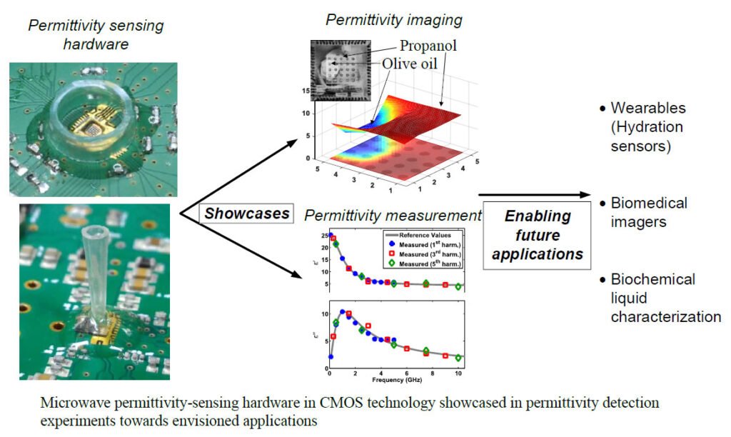
Circuits designed can act as the basic building block for a wide span of biomedical applications, ranging from wearables to permittivity imaging. Experimental results of manufactured prototypes demonstrate measurement noise reduction through bridge balancing, debye model parameter estimation of independent material with a 1.6% error using full frequency dataset and 5.3% in high energy efficiency mode, as well as image construction based on material permittivity differences.
×
Take Out Message
Take-Home Messages
- A compact, multi-purpose broadband architecture for integrated complex permittivity sensors, utilizing a patch element and a multiharmonic downconversion for fast and energy-efficient readout.
- The architecture can be embedded in systems performing GHz frequency range permittivity footprint measurement for material characterization as well as permittivity imaging.
- Applications range from traditional clinical and point-of-care scenarios to the increasingly emerging area of wearable devices. Examples include in-vivo tissue hydration monitoring, label-free malignant tissue inspection as an assisting tool in removal surgery, evaluation of drug penetration through skin and bloodglucose concentration measurement.
- The proposed sensor readout core has the smallest known area and is the first to demonstrate permittivity imaging capabilities at microwave frequencies.
A Versatile Magnetic Exposure System for In-Vitro, Ex-Vivo and In-Vivo Experiments Finalized to Therapeutic Applications in the IF Range
Elena Della Valle, Micaela Liberti, Francesca Camera, Alessandra Paffi, Stefania Petralito, Vincenzo Roncace, Costanza Burattini, Giorgio Aicardi, Francesca Apollonio.
Read Description
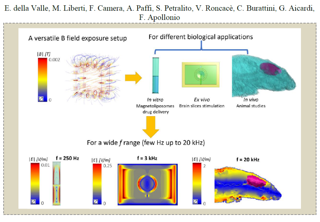
Laboratory experiments aimed at defining in vitro and in vivo outcomes are required, and reliable low intensity magnetic field exposure systems are needed. In the present study, we have performed the analytical and numerical design of a novel magnetic exposure system suitable for different biological applications, such as magnetoliposome drug delivery, ex vivo experiments on brain slices and in vivo studies. This system is be able to generate intensities of the order of mT in a frequency range from ELF to 20 kHz.
×
Take Out Message
Take-Home Messages
- A versatile magnetic exposure system able to reach intensities in the order of mT has been theoretically designed, with the aim of using it for different biomedical applications of low intensity magnetic fields, from a few Hz to 20 kHz.
- The biological applications for which this exposure system has been designed are very different: a cuvette for drug delivery applications; a chamber for ex vivo experiments on brain slices, and a rat phantom for in vivo animal studies.
- The system is designed to reach a magnetic field of 1.4 mT with a homogeneity of 95% in the volume between the coils where the target will be placed.
- The novelty of the proposed exposure system mainly relies on its versatility, which permits in vitro, ex vivo and in vivo laboratory experiments in a wide frequency range and with negligible thermal increase induce.